Hormones, insulin and blood sugar regulation

This subpage is the fourth part of the theory of Biotech Academy’s material on Diabetes.

You have now been introduced to some of the important organs associated with diabetes, including the digestive tract and pancreas. The pancreas, which produces and releases the hormones glucagon and insulin, among others, plays a major role in keeping blood sugar levels more or less constant. You have also read about the function of glucose in the body and how it is stored, broken down and used by the body’s cells. This section explains why many of these processes are dependent on insulin.

You will be able to read about how the different hormones described earlier affect each other and how together they regulate the body’s blood sugar levels and thus sustain our lives. You probably already know that even a short-term lack of oxygen to the brain will cause serious brain damage or even death, but did you know that the brain’s need for food in the form of glucose is just as important? Even a few minutes of glucose deprivation will cause unconsciousness and then death if glucose is not administered in time. After reading this article, you will know how our appetite is regulated and you will have learned about the key hormones related to satiety. You will get an explanation of how and when insulin is released and how blood sugar is regulated in a healthy individual. And last but not least, you want to know how insulin works when it is released into the bloodstream.

Our desire to eat is determined by habits and hormonal influences. The hormones leptin, which is produced and secreted from fat cells, and ghrelin, which is primarily produced and released from the stomach and duodenum, influence our behavior around food. The regulation of lactation occurs when hormones are released from the organs into the bloodstream and reach the brain, specifically the hypothalamus. This area of the brain is made up of many different nuclei with different functions, but here it should be described as a single unit that receives and sends signals.

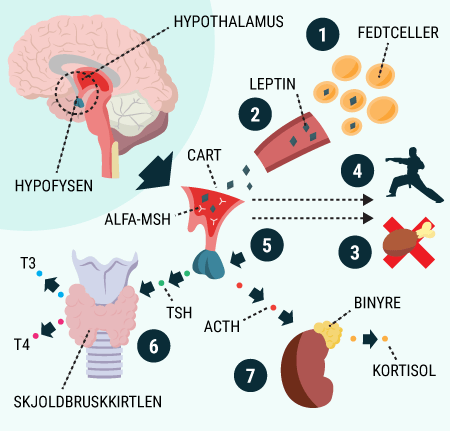
For years, homeostatic regulation of food intake has been studied, but it’s only recently that the understanding is coming into place. In 1994, a group of scientists managed to isolate a peptide hormone they called leptin, after the Greek word for ‘slim’. The peptide hormone leptin was found to signal to the brain that fat reserves in the body are normal. Leptin regulates our BMI by acting directly on the neurons (nerve cells) in the hypothalamus that decrease appetite and increase energy expenditure. A high leptin level in the blood will activate the leptin receptors, an alpha-melanocyte stimulating hormone (alpha-MSH) and cocaine and amphetamine regulated transcripts (CART) in the hypothalamus.

NAME ABBREVIATION FUNCTION
Alpha-melanocyte stimulating hormone Alfa-MSH Alpha-MSH is a hormone whose primary role is
contributing to the coloration of our hair and skin through a process called melanogenesis.
But it’s also important for our feeding and sexual behavior, among other things.
Cocaine and amphetamine regulatory transcripts CART CART is a peptide that plays a role in food intake, reward and stress.
Neuropeptide Y receptor NPY NPY is a receptor involved in the control of various behaviors including appetite, circadian rhythm and anxiety.
Agouti-related peptide AgRP AgRP is expressed together with NPY and works by increasing appetite and slowing metabolism.
Adrenocorticotropic hormone ACTH ACTH is the “adrenal cortex-directed hormone”. It is produced in the pituitary gland and released into the blood. ACTH’s function is to stimulate the adrenal cortex to release cortisol.
Thyroid-stimulating hormone TSH TSH is produced in the pituitary gland and released into the blood, where it stimulates the thyroid gland to release T4, which is converted to T3, actively stimulating metabolism.

Alpha-MSH and CART increase metabolism in the body. This is done by adrenocorticotropic hormone (ACTH) triggering cortisol release from the adrenal cortex and thyroid stimulating hormone (TSH) increasing thyroid activity. ACTH will therefore cause activity in the sympathetic nervous system. This activity will ultimately lead to reduced food intake (see figure 15).

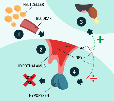
The opposite is true for low leptin levels in the blood. A low leptin level will stimulate other types of receptors, neuropeptide Y receptor (NPY) and agouti-related peptide (AgRP), in the hypothalamus. But the effect of NPY and AgRP on energy balance is the reverse of the effect of alpha-MSH and CART. NPY and AgRP will inhibit TSH and ACTH secretion, thus activating the parasympathetic nervous system. An activation of the parasympathetic nervous system will stimulate food intake behavior (see figure 16), which is why the NPY and AgRP receptors are also called the orexigenic peptides from the Greek word “appetite”.

Figure 15. This figure shows how leptin release from fat cells affects the body’s metabolism. 1) Fat cells produce and release leptin. 2) Leptin travels via the bloodstream to the hypothalamus, where the increased leptin level is detected by CART and alpha-MSH. 3) A signal is sent to the brain to inhibit food intake. 4) The sympathetic nervous system is activated. 5) A signal is sent to the pituitary gland to release TSH and ACTH. 6) TSH causes the thyroid gland to increase its activity and thus metabolism by releasing T3 and T4. 7) ACTH activates the adrenal glands to release cortisol, which increases metabolism.

Leptin is released from fat cells in the “saturated” body and travels with the blood to the hypothalamus. Here it stimulates alpha-MSH and CART to inhibit food intake, activate the sympathetic nervous system and cause the pituitary gland to release TSH and ACTH, which in the thyroid and adrenal glands will increase metabolism. Conversely, a starving body will cause decreased leptin release from fat cells. This lowered leptin level will be detected by NPY and AgRP, which will stimulate food intake and inhibit the release of TSH and ACTH, thus slowing down the body’s metabolism.

Figure 16. This is the body’s reaction to a reduced leptin level. 1) No leptin is produced and released from fat cells, lowering leptin levels in the blood. 2) NPY and AgRP detect the lower leptin level. 3) AgRP and NPY signal the brain to stimulate food intake. 4) NPY and AgRP signal the pituitary gland to inhibit the release of ACTH and TSH, ultimately leading to decreased metabolism.

The desire to eat, the eating process and the subsequent feeling of fullness can be divided into three phases:

  • The cephalic phase, where the sight and smell of food triggers physiological processes that activate the parasympathetic nervous system. An activated paraympathetic nervous system will initiate saliva production in the mouth and acid will be released in the stomach to break down food.
  • The gastric phase, where already initiated above mechanisms are greatly amplified as you start chewing, swallowing and filling your stomach with food.
  • The substrate phase when the stomach fills and the digested food moves through the intestines while nutrients are absorbed into the bloodstream.

It wasn’t until 1999 that the peptide hormone ghrelin was discovered, which is mainly produced in the stomach and released into the bloodstream when the stomach is empty. When ghrelin is injected into the blood vessels, it has been shown that this hormone is able to activate NPY and AgRP receptors in the hypothalamus, thus increasing appetite.

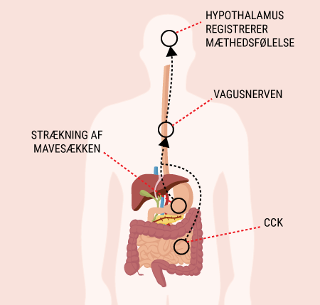
Figure 17. CCK and abdominal wall stretching both send satiety signals through the vagus nerve to the brain. The effect is synergistic, meaning the signals reinforce each other.

Gastric dilation and cholecystokinin
The stomach has both sensory and motor neurons. Sensory neurons carry signals from sensory organs to the brain, while motor neurons carry signals from the brain to the rest of the body. What these motor and sensory neurons have in common is that they primarily run along the vagus nerve. When the stomach wall stretches, sensory neurons detect a twitch and the motor neurons send a signal to the stomach to stop ghrelin release. A lack of ghrelin will then lead to a feeling of fullness, so we stop eating. In the duodenum , cholecystokinin (CCK) also plays an important role in food intake. Cholecystokinin is released from cells in the duodenum in response to intestinal stimulation from certain, especially fatty, foods. CCK’s primary role is to provide satiety, which also goes through the vagus nerve, so CCK and gastric dilation act synergistically (amplifying each other’s effect) to inhibit food intake (see figure 17).

NAME RELEASED FROM EFFECT
Leptin The fat cells (adipocytes) When leptin levels are high in the blood, it activates
sympathetic nervous system, metabolism increases and the hypothalamus signals the
stop food intake. When leptin levels are low, metabolism will slow down and
food intake is stimulated.
Ghrelin Primarily from the stomach and duodenum
but also in small amounts from the pancreas, gonads, lungs, and hunger
(jejenum)
An empty stomach releases ghrelin. When
ghrelin levels in the blood are high, receptors in the hypothalamus and
stimulates food intake.
Cholecystokinin (CCK) Enteroendocrine cells (producing cells in the
gastrointestinal tract) in the mucosa, in the duodenum and in the hunger intestine
(jejenum)
When the gut is stimulated – especially by fatty foods
CCK is released, whose primary function is to provide a satiety signal that is sent to
the brain via the vagus nerve. CCK works synergistically with abdominal wall stretching.


But how does insulin affect our blood sugar regulation? Read more in the next section.

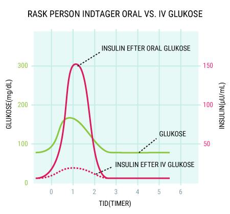
In “The Physiology and Anatomy of Diabetes”, you were briefly introduced to the secretion of insulin by the pancreas and its effects. The human body consists of 1014 cells and whether or not glucose can enter the cell depends on which GLUT transporter the cell has. There are a total of 12 different GLUT transporters in humans. The GLUT4 transporter is insulin-dependent, meaning that when the cell has a GLUT4 transporter, insulin must be present for the cell to take up the glucose. The GLUT4 transporter is primarily found in striated muscle (cardiac and skeletal muscle) and in adipose tissue. This means that these tissues are dependent on insulin to function. But the release of insulin is more complex than that and is influenced by multiple mechanisms. In a healthy person, blood sugar levels will usually be around 4-5 mmol/L after an overnight fast, but will rise after a meal. However, even with large and sugary meals, it will not rise above 11 mmol/L. If the healthy body detects a drop in blood sugar of just 20%, insulin release will be significantly lowered to avoid hypoglycemia (low blood sugar) at all times. In this way, it will be food intake or fasting that has the greatest influence on blood sugar changes in healthy individuals. However, it’s not just blood sugar that determines how much insulin is released. It also matters whether the glucose is taken orally (by mouth) or injected directly into the bloodstream. If the glucose is taken orally, it will cause a much higher insulin secretion, as incretins such as gastric inhibitory polypeptide (GIP), glucagon-like peptide-1 (GLP-1) and CCK are secreted in the gut upon ingestion and enhance insulin release (see figure 18). The incretins are particularly exciting in research, as the mechanism can be used to develop medication for diabetics.

Figure 18. The graph illustrates that when a healthy person consumes a meal with glucose, blood sugar (the green graph) will rise slowly. In response, the beta cells in the pancreas release insulin, causing the concentration of insulin (the red graph) to increase significantly. If the glucose is given into the bloodstream, insulin will rise slightly but not nearly as much as with oral intake (the dotted red graph). This is due to the incretin effect, which can be exploited in the pharmaceutical industry for the development of diabetes drugs.
Please note that blood sugar in this figure is expressed in mg/dL. A slightly simplified way to convert blood sugar from mg/dL to mmol/L is: mmol/L = mg/dL/18
The maximum blood sugar in this figure is about 170 mg/dL, which corresponds to a blood sugar of about 9.4 mmol/L.

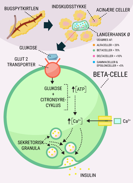
But how can glucose affect the release of insulin in the beta cell of the healthy individual?

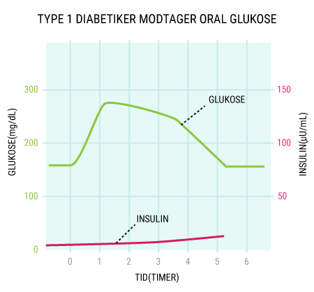
Figure 19. Type 1 diabetics take oral glucose.

The aforementioned hormones insulin and glucagon, which are produced in and released from the pancreas, have opposing effects and are responsible for up- and down-regulating blood sugar levels respectively. When blood sugar levels are high, insulin promotes anabolic processes in muscle, liver and adipose tissue by stimulating glycogen, protein and fatty acid synthesis. At the same time, insulin ensures that glucose can be transported across the cell membrane via GLUT transporters, thereby both nourishing the cells and lowering blood sugar levels.

Glucagon works in the opposite direction by initiating glycogenolysis (the breakdown of glycogen into glucose), which can then increase blood sugar levels. In addition, glucagon stimulates gluconeogenesis (new glucose formation) and lipolysis (breakdown of fat into free fatty acids) to provide available energy and inhibits glycolysis (breakdown of glucose) to maintain normal blood sugar levels. By constantly measuring and regulating and then releasing glucagon and insulin, the body can ensure that blood sugar levels are neither too high nor too low. When blood sugar levels rise, insulin is released, while glucagon is released when blood sugar levels drop.

Figure 20. This shows the mechanism behind insulin release in response to increased blood sugar. Glucose reaches the beta cell in the pancreas via a GLUT2 transporter. As blood sugar rises, increased intracellular ATP will cause increased calcium influx into the cell. The increased amount of calcium will influence the secretory granules to release insulin from the beta cell.

In addition to blood sugar, which is the main regulator of insulin secretion, we also have hormonal regulation exerted by adrenaline and noradrenaline, which bind to two different receptors on beta cells. Binding to one receptor, the alpha receptor, inhibits insulin secretion, while the other receptor, the beta receptor, increases insulin secretion. Overall, however, the alpha receptor signal is the strongest and the result is reduced insulin secretion when we have large amounts of adrenaline and noradrenaline circulating in the blood. This is smart because large amounts of adrenaline or noradrenaline are linked to stress in the body, for example during “fight and flight”. When the body prepares to fight or flight and adrenaline and noradrenaline are released, it causes many reactions in the body. Examples of this are increased pumping function in the heart, contraction of smaller vessels in the body to increase blood pressure and the breakdown of brown adipose tissue to generate heat for the body. Of course, all of these processes require energy, so glucose and therefore energy must be released.

The sympathetic and parasympathetic nervous system
The nerve cells in the body that are not part of the brain, brainstem and spinal cord are collectively known as the peripheral nervous system. The peripheral nervous system is divided into sensory, motor and autonomic parts. The autonomic nervous system (ANS) regulates functions in the body that are unconscious to us, such as the heart muscles, our internal organs and the contraction or dilation of blood vessels. The ANS can be subdivided into the sympathetic nervous system, sympatheticus, and the parasympathetic nervous system, parasympatheticus. In simple terms, you could say that sympathetic nervousness is activated in stressful situations, such as when we are exposed to danger, the so-called “fight or flight” response. Similarly, the parasympathetic nervous system can be considered responsible for the processes that take place in the body when we rest, such as digestion, sexual desire, urination and defecation. An old mnemonic to separate sympathetic and parasympathetic nervous system is “you
p
isser and
p
arer with
p
arasympatikus” (but you probably shouldn’t say that out loud in the exam). Both the sympathetic and parasympathetic nervous system have receptors to which ACTH binds. In the sympathetic nervous system, ACTH acts as a neurotransmitter by releasing norepinephrine and epinephrine, which stimulate the so-called adrenergic receptors in the peripheral target tissue of the sympathetic nervous system. Similarly, ACTH binding to receptors in the parasympathetic nervous system will cause the release of more ACTH, which stimulates the muscarinic receptors in the target organs of the parasympathetic nervous system.

The opposite process takes place when the “danger has passed” and the body is no longer in a stressed state. When the parasympathetic nervous system is activated, insulin secretion increases again (if blood sugar levels allow) and the anabolic phase of rebuilding glycogen stores resumes (see figure 21).

The final form of insulin stimulation is the previously mentioned incretins GIP and GLP-1, both of which are secreted in the small intestine after glucose ingestion. They stimulate insulin release and are therefore important players in regulating the amount of insulin in the blood. If the overall production of insulin is insufficient and we are unable to absorb enough glucose from the blood, the disease diabetes mellitus occurs.

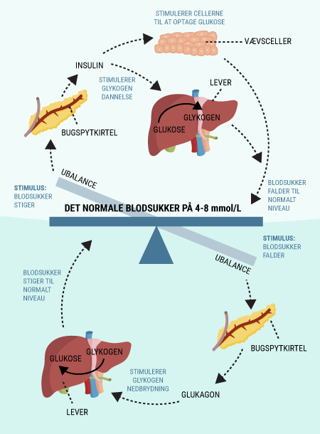
Figure 21. Blood sugar regulation in a fast individual. The entire blood sugar regulation in a healthy individual can be described as a delicate scale that will constantly try to achieve balance to maintain normal blood sugar levels. When blood sugar levels rise, insulin will be released, stimulating cells to take up glucose and the liver to convert glucose into glycogen, known as glycogenesis. That way, blood sugar levels will return to normal. When blood sugar levels drop, the pancreas is stimulated to release glucagon, which causes the liver to convert glycogen into glucose, known as glycogenolysis. Once glycogenolysis takes place, the glucose will be released into the blood and blood sugar levels will return to normal.

As mentioned earlier, insulin works by increasing anabolic processes in muscle, liver and fat tissue, but the way it does this in different tissues is slightly different. In general, insulin binds to insulin receptors on liver cells(hepatocytes), muscle cells (myocytes ) and fat cells (adipocytes). After insulin binds to the insulin receptor, different intracellular processes (activities inside the cell) are initiated depending on the cell. Basically, insulin will increase the build-up of the different energy stores we have in the body, for example by affecting glucose uptake and storage, so that glucose can be stored as glycogen. Insulin will help increase protein synthesis and inhibit proteolysis, the breakdown of proteins. High plasma insulin will increase the formation of triglycerides in the liver, which can then store the triglycerides or send them through the blood to fat and muscle tissue, which can later, if needed, oxidize the triglycerides and release energy. While plasma insulin is high, it’s the carbohydrates that need to be used and therefore lipolysis and lipid oxidation (fat breakdown) are inhibited.

But what exactly does insulin do that is so important for sustaining life?

As mentioned earlier, it is cardiac and skeletal muscle as well as fat cells that rely on GLUT4 transporters. Together, these three cell types account for about 2/3 of all cells in the human body, and while muscle cells are responsible for our movement and breathing, fat cells are important because they can store the energy we consume for later use. A high level of insulin in the blood will cause an insulin molecule to bind to the insulin receptor in, for example, a muscle cell, leading to a change in the intracellular part of the insulin receptor. This is followed by a series of intracellular processes that upregulate the amount of GLUT transporters and inactivate the enzyme glycogen synthase kinase, resulting in the formation of more glycogen in the cell (Figure 22).

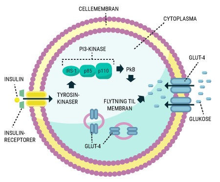
Figure 22. The figure shows how insulin enables glucose uptake in, for example, a muscle cell. When insulin binds to the insulin receptor, its structure changes and tyrosine kinases are activated. The tyrosine kinases activate the enzyme PI3-kinase, which catalyzes the formation of PkB. PkB then sends GLUT4-containing vesicles to the cell membrane. A higher number of GLUT4 transporters in the membrane increases the cell’s glucose uptake, thus providing energy to the cell while lowering blood sugar levels.

In-depth explanation of how insulin works
A high level of insulin in the blood will cause an insulin molecule to bind to the insulin receptor in, for example, a muscle cell, leading to a change in the intracellular part of the insulin receptor. This change in the insulin receptor leads to a cascade reaction that starts with the activation of the tyrosine kinases. The tyrosine kinases can then phosphorylate the enzymes MAP kinase, which initiates cell growth and gene expression, and PI3 kinase, whose main function is shown in figure 22, namely to send GLUT4 transporters to the cell membrane. This is done by the enzyme PI3K catalyzing the reaction:

PIP2 + ATP -> PIP3 + ADP

PIP3 will then activate protein kinase B (PkB), which facilitates vesicle fusion. In this way, GLUT4 transporters, which otherwise reside in vesicles in the cell, are transported to the cell membrane, allowing glucose to enter the cell (see figure 22). In addition, PkB also inactivates glycogen synthase kinase (GSK), ultimately allowing more glycogen to be formed in the cell. This is related to the increased number of GLUT4 transporters and thus increased amount of glucose in the cell, which needs to be stored in the form of glycogen.


Liver cells (hepatocytes)
The above mechanism with GLUT4 transporters takes place in striated muscle and fat cells, but in liver cells the process is slightly different. This is because liver cells do not have GLUT4 transporters, which are dependent on insulin, but instead have GLUT2 transporters.

When insulin rises, the liver is forced to take glucose from the blood and either convert it to glycogen through glycogenesis or break it down into pyruvate. This substance is a necessary building block for storing glucose as fat. High blood sugar will increase the formation of fat, triglycerides, in the liver. The liver then has two options with the newly formed fat; it can either store the triglycerides in the liver or – if there is a lot of fat – send them through the bloodstream to fat and muscle tissue respectively. In fat and muscle tissue, the fat can be broken down by oxidation, so the energy can be used when needed. However, insulin also works by slowing down the oxidation of fat, which normally releases ATP for burning. In doing so, insulin forces the liver, as well as other tissues, to burn the carbohydrates and reduce blood sugar levels.

You have now learned how our food intake is regulated by leptin, which is released from fat cells and affects the hypothalamus. When leptin is high, the sympathetic nervous system will be activated and food intake is inhibited, while a low leptin level will activate the parasympathetic nervous system and stimulate food intake. You’ve also learned that blood sugar levels in healthy individuals are always fairly stable, and that this is due to a balanced glucagon and insulin release from the pancreas. You now know that taking glucose orally rather than intravenously will cause plasma insulin to rise much more because the incretins GIP, GLP-1 and CCK are released when sugar is taken by mouth. This is important to remember when you later read about the pharmaceutical industry and its production of medicines. Here, biological processes are harnessed to create the most optimal medicine. There are a total of 12 different GLUTs, and you have learned about GLUT2, which is found in the pancreas and liver, and GLUT4, which is found in striated muscle (skeletal and cardiac muscle) and adipose tissue. The main difference between GLUT2 and GLUT4 is that the latter is insulin dependent. In practice, this means that striated muscle and fat tissue can only function when pancreatic beta cells produce and release insulin. Finally, you will have briefly understood the processes that occur in liver, muscle and fat cells when plasma insulin is high after glucose ingestion. You now know that the body will utilize the carbohydrates to convert glucose into glycogen, to form triglycerides and to increase protein synthesis, so that all energy stores are full in case the body needs energy later on.