CRISPR/Cas9 – The genetic engineering revolution

Summary: CRISPR technology comes from the immune system of prokaryotes, where it acts as protection against viral attacks, for example. CRISPR is an abbreviation of Clustered Regularly Interspaced Short Palindromic Repeats, which describes the genomic structure of the prokaryotic immune system. CRISPR associated (Cas) proteins are the functional units of the immune system. The most commonly used type of Cas9 in genetic engineering comes from the bacterium Streptococcus pyogenes.


Everything that can be defined as life has one unique thing in common. All the information that forms individual organisms is chemically stored in a macromolecule that functions in the same fundamental way in man and all other organisms. DNA is the molecule that makes up our genetic information through its genetic code. It is of enormous importance that life shares the foundations of this genetic code, as it means that it is possible to take information from one organism and use it in another. Genetic engineering makes it possible to move genes around and control their activity, so that we can actually control the shape of life. Genetic engineering can be used to create untold amounts of genetic combinations that, for example, can give us better food, change the properties of organsims, be used for the production of medicine or even cure diseases. It is only the imagination and technology that limits the possibilities – fortunately, technology has come a long way!

One of the latest technologies is CRISPR/Cas9, which has great potential because it provides the opportunity to cut DNA with enormous precision and efficiency, in an easier and cheaper way. This technology is based on one protein called Cas9. The protein is controlled by a piece of RNA called guide RNA (gRNA), which gives the protein its property to accurately identify DNA sequences, bind to them, and cut them by forming precise double-strand breaks in DNA. The reason why Cas9 has so much potential is that the protein can easily be reprogrammed to target new DNA sequences by replacing the gRNA that controls the protein. Cas9’s precise formation of double-strand fractures allows, for example, the insertion of DNA sequences into very specific positions with which new gene functions can be obtained. One can also form mutations that can deactivate specific genes and thus remove specific traits from organisms.

The CRISPR technology originates from the immune system of prokaryotes, where it acts as protection against viral attacks, for example. CRISPR is an abbreviation of C lustered Regularly Interspaced Short Palindromic Repeats, which describes the genomic structure of the prokaryotic immune system. CRISPR asassociated (Cas) proteins are the functional units of the immune system. The most commonly used type of Cas9 in genetic engineering comes from the bacterium Streptococcus pyogenes.

CRISPR/Cas9 is a magnificent example of a biotechnological tool developed from nature’s own functional mechanisms.

The basic theory will provide a good basic understanding of the molecular function of CRISPR/Cas9 and how we can exploit it for gene modification. Then the different cases can be read independently of each other.

Theory:

Access the theory material by pressing the buttons below.

Cases and tasks:


  • Case 1 - The Origins of a Prokaryotic Immune System

    The origin of CRISPR/Cas9 – a prokaryotic adaptive immune system

    CRISPR/Cas systems are prokaryotic immune systems designed to defend bacteria and archaea against invasive genetic material. This could be, for example, plasmids, which are small ring-shaped DNA sequences that can affect the prokaryote’s genome, or viruses that take over the cell functions of the prokaryote so that the virus can reproduce itself. In general, it can be said about invasive genetic material that it tries to alter the cell’s DNA, with which it poses a potential threat to the genome.

    CRISPR/Cas systems generally consist of two components, a DNA section called the CRISPR region, found in the prokaryote’s own genome, and the functional Cas proteins that move around the cytoplasm. For example, in a viral attack on a prokaryote, viral DNA will be injected into the cell, which must be destroyed quickly before it takes over the cell’s functions and kills it. Here, the CRISPR/Cas system is quickly activated, whereby Cas proteins are sent out into the cell’s cytoplasm to search for the invasive DNA. As soon as the foreign DNA is located, it is cut into pieces by double-strand fractures and then broken down so that it no longer poses a threat.

    The system is said to be adaptive, as an infection collects a small sample of the invasive DNA, which is stored in the CRISPR region and can be used to more quickly recognize future infections of the same type. It is these fragments that are used to make gRNA for Cas9, so that the protein can recognize the invasive genetic material. It is these fragments that make up the immunity of the system and they can be inherited by the offspring of the bacteria, giving the new bacteria the same immunity that previous generations have acquired.

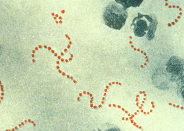
    There are an incredible number of different variations of CRISPR/Cas systems, with different proteins and mechanisms of action that vary between prokaryotes. The bacterium Streptococcus pyogenes contains two types of CRISPR/Cas systems, namely type I and type II. Little is known about the mechanism of operation of the Type I system and whether it is active at all. The Type II system, on the other hand, is very well studied and was used to develop the much talked about genetic engineering tool, CRISPR/Cas9. S. pyogenes is depicted in Figure 14. To understand how the genetic engineering tool was discovered, one can look at how the original immune system works, as the functional mechanisms are very similar. This now takes a closer look at the Type II CRISPR/Cas system in S. pyogenes.

    Figure 14. Streptococcus pyogenes is the bacterium where the most commonly used CRISPR/Cas9 originates. The red dots are individual bacteria that together form chains.

    Source: Centers for Disease Control and Prevention, United States Department of Health and Human Services.

    The structure of the type II CRISPR/Cas immune system

    CRISPR region

    The CRISPR region is the immune system’s information hub, where all information is stored and where the Cas proteins are emitted from or reported to. CRISPR is short for C lustered Regularly Interspaced Short Palindromic Repeats. The name very accurately describes the characteristic structure of the CRISPR region of the prokaryotic genome; repeats of small DNA sequences, called repeats, that lie together at the same distance from each other. These are 32 base pairs long. Repeats are palindrome, meaning they are read identically both from left to right on one string and in the opposite direction on the complementary string.
    Between each repeat, there is a DNA sequence of extragenomic origin, which means that the sequences do not originate from the prokaryote’s own genome. These are called spacers and are roughly 35 base pairs long fragments of invasive DNA from previous infections. They are derived from bacteriophages (viruses), plasmids or other foreign DNA. These acquired spacer fragments are used as a library of previous infections, so that the system can easily and quickly identify DNA from infections that the cell has experienced in the past. Each spacer thus corresponds to a previous infection of the bacterium. The spacers can be transcribed into RNA and converted into gRNA, which is the form in which they are used by Cas proteins.

    In the CRISPR region, a leader is held, which is a larger regulatory DNA sequence that determines the transcription of spacers into usable gRNA and controls the integration of new spacers. Furthermore, the Cas genes are available, which are very important for the system as they code for the Cas proteins. They are located near the CRISPR region of the genome. See Figure 15 for the structure of the CRISPR region and the cas genes for the Type II CRISPR/Cas system in S. pyogenes.

    Figure 15 – Structure of the genetic region of the type II CRISPR/Cas system in S. pyogenes. The CRISPR region contains all the important information about previous infections, in the form of various spacers that sit between repeats. The region contains a leader, which is a DNA sequence that regulates the integration and transcription of spacers. The four cas genes encode the Cas proteins that exercise the functions of the immune system. The nearby tracrRNA gene is used in the formation of gRNA for Cas9.

    The Cas proteins

    CRISPR asassociated (Cas) proteins are the functional units of the immune system – they are the ones that do the work. Their function is to maintain the activity of the immune system and take care of the structure of the CRISPR region, which is arranged by different proteins with different roles. Some Cas proteins collect information about new infections, in the form of the spacers, which they deliver to the CRISPR region, while other Cas proteins are emitted from the CRISPR region with this information to eliminate the infection threats. Cas proteins can be controlled by gRNA, similar to the DNA sequence that the system wants them to work on. Specifically for the Type II CRISPR/Cas system in S. pyogenes, the following Cas proteins are present:

    Cas1 – collects new spacers for the CRISPR region together with Cas2.

    Cas2 – collects new spacers for the CRISPR region together with Cas1.

    Cas9 – detects and eliminates invasive DNA by forming double-strand breaks in the DNA. The protein is controlled by guide RNA (gRNA), which originates from the spacers of the CRISPR region.

    Csn2 – involved in the acquisition of new spacers for the CRISPR region.

    In addition, there is one important protein that is necessary for the functioning of the system:

    RNase III – processes RNA transcribed from the CRISPR region and forms the finished gRNA, which is used for the control of Cas9.

    Function of the Type II CRISPR/Cas immune system

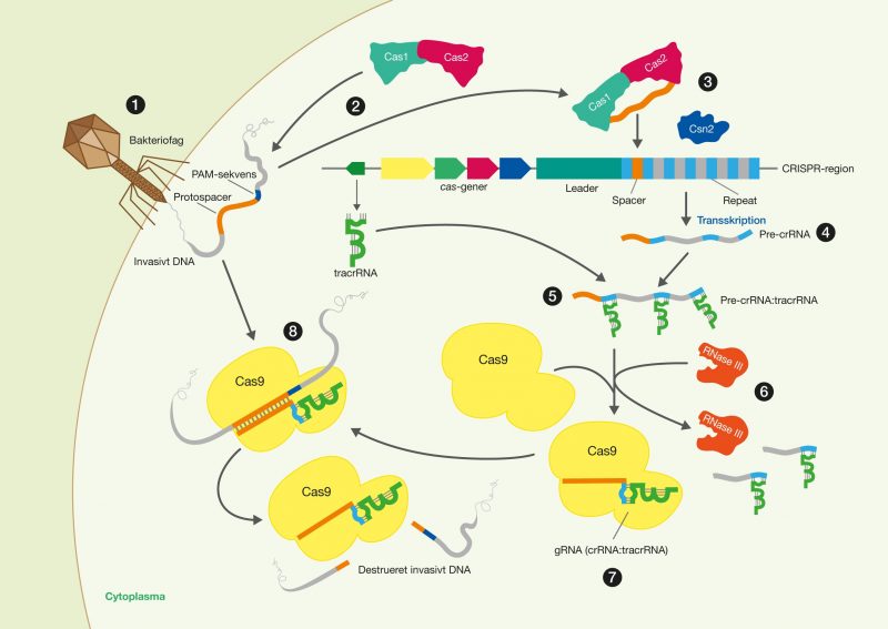
    The following section will describe the mechanism of operation of the type II CRISPR/Cas immune system, exemplified by bacteriophage attacks on the bacterium Streptococcus pyogenes. The process is described step by step, as shown in Figure 16.

    2.2_crop

    Figure 16. Overview of the type II CRISPR/Cas immune system response to a bacteriophage infection in S. pyogenes. The individual steps are numbered corresponding to the detailed description below.

    Acquiring new immunity: adaptation

    The CRISPR/Cas immune system is adaptive, as the system can acquire immunity against new invasive genetic material. This means that the system can adapt to new threats that have not infected the cell before. The following steps describe how the DNA from a bacteriophage is converted into immunity to the bacteriophage itself, by the CRISPR/Cas system in Streptococcus pyogenes.

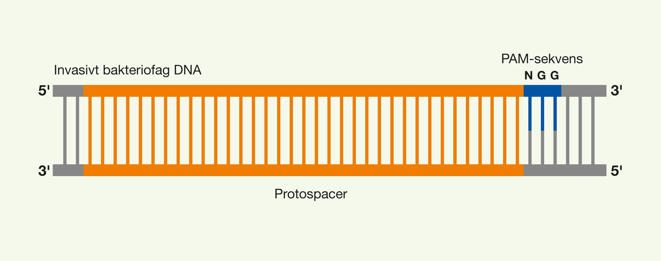
    1. A bacteriophage binds to a surface protein on the surface of the prokaryote, thereby starting the infection. The bacteriophage shoots its invasive DNA into the cytoplasm of the prokaryote, which is now an acute threat to the cell as the bacteriophage DNA tries to take over the cell’s mechanisms of action. On the invasive DNA there is Protospacer Adjacent Motifs (PAM), which defines the area of the invasive DNA that the CRISPR/Cas system can turn into a spacer. This area is called a protospacer. PAM sequences are apparently randomly placed, making the protospacer a randomly selected part of the invasive DNA. The PAM sequence for S. pyogenes is 5′-NGG-3′. This means that wherever 5′-NGG-3′ is seen in the invasive DNA, a potential protospacer is available. The interesting part of the invasive DNA can be seen in Figure 17.

    Figure 17. A sample of the invasive DNA and its location of the protospacer and the essential PAM sequence, 5′-NGG-3′, recognizable by Cas proteins.

    2. The CRISPR/Cas system reacts to the infection and Cas proteins examine the invasive DNA. The Cas proteins Cas1 and Cas2 form a complex that may also interact with Cas9 and Csn2. This Cas1-Cas2 complex investigates the invasive DNA and is responsible for the selection of protospaces from recognition of PAM sequences, but this is done by an as yet undetermined mechanism for S. pyogenes. The protospacer is cut free from the invasive DNA by the Cas1-Cas2 complex and transported to the CRISPR region.

    3. The Cas1-Cas2 complex also acts as an integrase, inserting the newly acquired approximately 35 base pair spacer into the CRISPR region. Integration of a spacer must be done very accurately, so as not to destroy the CRISPR region. The Cas1-Cas2 complex integrates the spacer depending on the leader sequence and when recognizing repeats. The spacer is inserted between two repeats at the end of the leader sequence. Once the spacer has been incorporated into the CRISPR region, it provides potential immunity to the bacteriophage that attacked the cell. Immunity is defined by the specific sequence of the spacer, as this could be used to quickly identify a new attack of the same type of bacteriophage. In order to exercise the immunity that the spacer provides, the spacer must first be reformulated into usable gRNA for Cas9. Thus, another mechanism of the system must be activated.

    Formulation of immunity: CRISPR expression and formation of gRNA

    Cas9 is the protein that identifies invasive DNA and then destroys it. In order for Cas9 to recognize the threatening invasive DNA, it must formulate the corresponding newly acquired spacer in a way that allows it to be used practically. The spacer will be formulated as part of gRNA, which guides Cas9 towards the invasive DNA. Remember that gRNA is the interchangeable part of Cas9 that allows the protein to recognize various specific sequences, bind to them, and destroy them by cutting them. In bacterium, gRNA consists of two pieces of RNA, namely crRNA (CRISPR-RNA) and tracrRNA (trans-activating CRISPR-RNA). The following steps describe the formation of gRNA from the newly acquired spacer from the CRISPR region.

    4. The first step for the formation of gRNA is transcription of the CRISPR region containing the given spacer. This is believed to be promoted by the leader sequence. The transcription forms a longer piece of RNA consisting of a longer series of spacers and repeats. This RNA is called pre-crRNA.

    5. Near the CRISPR region, tracrRNA is transcribed in order to interact with pre-crRNA. This tracrRNA binds to the repeat segments of pre-crRNA, forming a pre-crRNA:tracrRNA complex.

    6. The formed RNA complex recruits the RNA-specific nuclease RNase III, which cleaves the RNA complex into several crRNA:tracrRNA complexes, in the presence of Cas9. The resulting RNA complexes consist of one piece of tracrRNA and one specific piece of crRNA, which actually make up finished gRNA.

    7. The formed gRNA with the spacer from the bacteriophage is now absorbed by Cas9, so that the protein becomes active. This allows Cas9 to search for the invasive DNA and destroy it before it’s too late. The crRNA portion contains a 20 nucleotide long portion of the spacer used by Cas9 for the identification of invasive DNA. The tracrRNA part plays a structural role that allows Cas9 to bind the gRNA and thereby use it.

    Exercising immunity: Cas9 activity

    The activated Cas9 now monitors the cell for bacteriophage attacks of the same type, by searching the cytoplasm for the bacteriophage DNA. The presence of the PAM sequence is a necessity for Cas9 to recognize the bacteriophage DNA, and thus it primarily searches for these. Each time Cas9 finds a PAM sequence, the protein tests whether the gRNA’s 20 nucleotides spacer portion matches the rest of the DNA sequence. Since PAM sequences are missing in the CRISPR region, it is ensured that Cas9 cannot recognize the spacer sequences of the CRISPR region and thus cannot cut them. The requirement for the PAM sequence thus serves as a security for the bacteria’s own CRISPR region, thus avoiding autoimmunity in the CRISPR/Cas system.

    8. When Cas9 meets the DNA of the bacteriophage, it will bind to the PAM sequence and the DNA of the bacteriophage will match the newly formed gRNA, since the two sequences are similar. This is because the bound gRNA in Cas9 is based on this very sequence, which made up the protospacer in the beginning. This gRNA DNA binding causes Cas9 to bind to the DNA strand. Since Cas9 has now identified the bacteriophage’s DNA, Cas9 makes a double-strand break, causing it to decompose. Cas9 lets go of the cleaved DNA and is then ready to search further.

    In this way, S. pyogenes is protected by the CRISPR/Cas system from bacteriophage attacks and the same mechanism is seen in plasmids or other genetic elements.

    Background history of the development of CRISPR/Cas9


    Cas9’s predecessors – Zinc-fingers and TALENs

    The idea of modifying genes is not new, and CRISPR/Cas9 has several predecessors. The previous genetic engineering tools, Zinc-fingers and TALENs (transcription activator-like effector nucleases), are made up of single protein units that can recognize a few nucleotides. These protein units can be composed into larger complexes so that they can recognize longer sequences. These protein complexes can be attached to a nuclease, which can then make a double-strand break in DNA. Zinc fingers can only recognize 3 nucleotides and TALENs only 1 nucleotide, so it takes a lot of work to build a protein complex that can give the same effect that is easily achieved by CRISPR/Cas9.
    A major advantage of Cas9 is that it is just a single protein that does not need any modifications every time a new sequence is hit, because it uses RNA-based identification rather than protein-based identification. Since you only have to replace the controlling gRNA to vary which sequence you want to hit, it is much easier than building new protein complexes. Therefore, CRISPR/Cas9 is lighter and cheaper than before.


    The development of CRISPR/Cas9

    The development of the genetic engineering tool CRISPR/Cas9 is a prime example of biotechnology. As with Alexander Fleming’s discovery of penicillin in 1928, and so many other biological discoveries, it often all starts by chance.
    The mysterious repetitive CRISPR DNA sequences have been known since Yoshizumi Ishino and his research team accidentally discovered them in 1987, in connection with some other genetic work. However, their function remained unknown until after the turn of the millennium, when various research groups began to investigate the CRISPR sequences. The sequences only became more mysterious when it was discovered in 2005 that they did not originate from the organisms themselves, but originated from viruses and plasmids. However, it was this discovery that led the researchers to hypothesize that the CRISPR sequences played a role in a prokaryotic immune system. Their function was experimentally confirmed in 2007 by a research team from the food company Danisco, who tried to vaccinate their bacterial strains against viruses and thus turn them into stronger production organisms. After this, the functional mechanism of the CRISPR/Cas immune system began to be clarified through various studies, which gave the starting point to the race to use the system for genetic engineering purposes. In 2012, some research groups had simultaneously proved that systems from Streptococcus thermophilus and Streptococcus pyogenes could be modified and constitute genetic engineering tools. These discoveries sparked the great patent war between the University of California, Berkeley, and the Broad Institute. This is an incredibly complex and heated conflict over who can call themselves the rightful owner of the CRISPR/Cas9 technology.

    In general, it can be said that the researchers’ curiosity led to the studies of the function of the CRISPR sequences, which gave rise to the discovery of Cas9. Studies of the function and structure of Cas9 in its natural environment revealed its great potential when it was discovered that the protein could both recognize and cut DNA sequences. The exploration of the formation and function of gRNA is absolutely central, as it was discovered that it was this element that was the key to the control of Cas9. It is based on this knowledge that Cas9 has been made programmable with synthetically produced sgRNA.

    The development of CRISPR/Cas9 tells the story that biotechnology is often about discovering and exploring the fundamental tools that nature itself has created and then figuring out how we can use them for beneficial purposes.

    Who knows what else is out in nature’s vast unexplored landscape?

  • Case 2 - Gene regulation with CRISPR technology

    Gene expression control with CRISPR technologies – CRISPRi and dCas9

    Genes encode many different properties, depending on the function of the proteins that the genes encode. In order for the properties to be expressed in an organism, the genes must first be transcribed. Thus, one can control the expression of properties by controlling the transcription of the genes.
    Other CRISPR/Cas9-based tools have been developed than just the natural Cas9. These also take advantage of the precise binding that Cas9 can perform in combination with sgRNA, but offer options other than double-strand breaking for genetic modification. They all are varieties of the Cas9 protein, which has been modified. It is possible to use the Cas9 protein to control gene regulation. By converting the Cas9 protein into an inactive form, a protein is formed that simply binds specifically to DNA sequences depending on the given sgRNA. This type of Cas9 is called dead Cas9 (dCas9) as it cannot make double strand breaks in DNA. dCas9 is formed by inactivating the Cas9 endonucleases, RuvC and HNH, with point mutations in the Cas9 gene, so that these cannot cut into DNA strands. dCas9 allows you to control the activity of genes by making it bind to specific sequences. This gene control method is called CRISPR interference (CRISPRi). With this method, it is possible to control whether genes should be expressed or inhibited without editing the DNA sequences themselves. CRISPRi does not alter the genetic information, but controls its transcription. This has its advantages, since the gene functions can be regulated without making permanent mutational changes in the DNA sequence, which can have serious consequences for the organism. Important advantages, then, are that the manipulation is reversible and non-mutational. The mechanism is similar to the normal function of Cas9, as dCas9 identifies DNA sequences with the given gRNA and from a PAM sequence, which causes a binding formation to the DNA sequence.


    Gene silencing with dCas9: repression

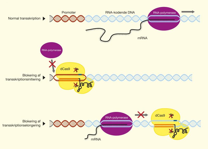
    It is possible to effectively reduce expression of a very specific gene if you can block the RNA polymerase, which transcribes DNA into mRNA. By using dCas9 and a piece of sgRNA, which is designed to recognize the same place as the RNA polymerase binds, a physical blockage of the RNA polymerase can be achieved. This is called gene silencing, as the expression of the gene is suppressed. There are several approaches to gene silencing with dCas9, and their effectiveness depends on where on the gene you hit.

    3.1_crop

    Figure 18. CRISPRi gene silencing with dCas9. At the top is the structure of the gene, with the promoter and the RNA-coding DNA. The RNA polymerase is seen in progress with the transcription. The blocking of transcription initiation shows how dCas9 settles in the promoter and blocks binding of the RNA polymerase, thus preventing the transcription of the gene. The blocking of the transcription longation shows how dCas9 settles in the middle of the gene and blocks the movement of RNA polymerase down the DNA strand, which also shows an inhibition of the transcription. Thus, the expression of the gene can be inhibited.

    Blocking transcription initiation – preventing the start of transcription. For the most optimal effectiveness, dCas9 is at the starting point of the transcription, the promoter. This will block the binding of the RNA polymerase itself, thus preventing the start of transcription. The blockage is independent of which DNA strand dCas9 binds to, as binding to the coding strand and the template strand both prevent binding of the RNA polymerase and thus provide equal repression of the gene. See Figure 18 for an illustration of transcription initiation blocking.

    Blocking transcription elongation – preventing execution of the transcription. dCas9 can also bind in the middle of the gene’s RNA-coding sequence, blocking the movement of RNA polymerase along the strand as it transcribes the gene. The effectiveness of this depends on which string dCas9 sits on. To achieve high effectiveness of repression, sgRNA must be designed so that dCas9 binds to the coding DNA strand and not to the template strand. See Figure 18 for illustration of blocking transcription elongation.

    As with traditional genetic modification with Cas9, multiplexing can be used, where several pieces of sgRNA can be used together with dCas9 to block several genes at once. It is also possible to block the expression of one gene extremely effectively by having multiple sgRNA that target the same gene but in different places.

    CRISPRi gene silencing for modification of Escherichia coli

    Bacteria can have an amazing variety of structures, shapes and sizes, which is referred to as their morphology. When describing the morphology, it is about the phenotype of the organism. The variations range from tiny spirals to large round bacteria visible to the naked eye (e.g. Thiomargarita namibiensis). Like all their other characteristics, their morphology depends on the genetic information stored in their DNA. This means that with the help of CRISPR technology, you can change the morphology of the bacteria, which can be beneficial, for example, when using the bacteria for production.

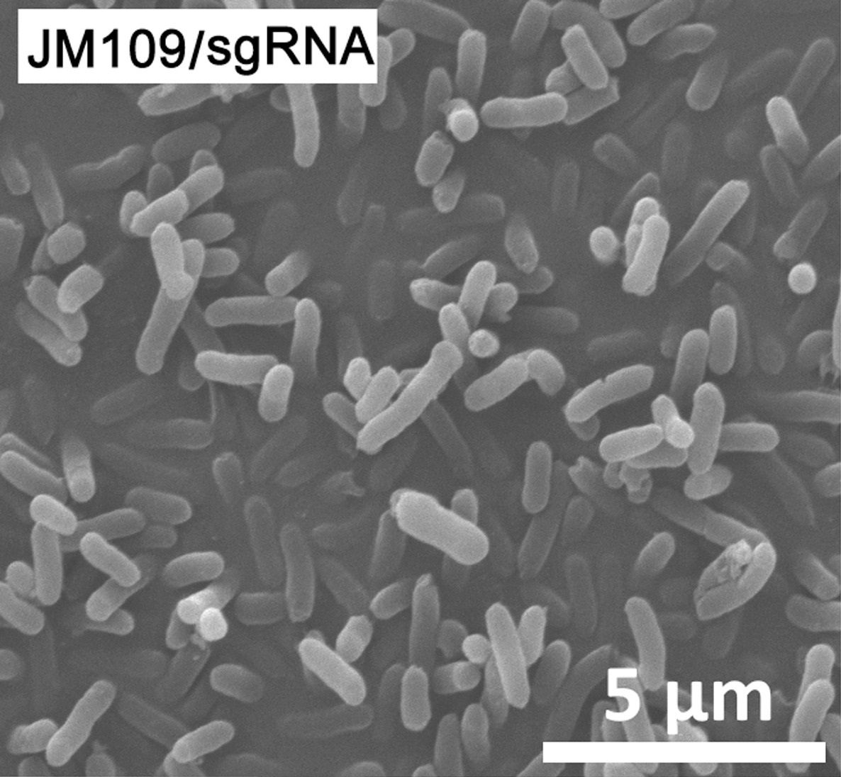
    A closer look will now be taken at how a research team tested CRISPRi to regulate the expression of genes that determine the morphology of Escherichia coli and thus be able to control their appearance. The researchers wanted to achieve larger E. coli bacteria, as these can increase the production of bioplastics due to a larger cell volume for the accumulation of the bioplastic. The normal morphology of E. coli is a slightly elongated rod shape, which can be seen in Figure 19. The picture shows the negative control from the CRISPRi trial, which means that the bacteria deliberately contain dysfunctional sgRNA, with which no genetic changes have been performed in these bacteria. They have the wild-type phenotype.

    Figure 19. The natural morphology of E.coli. This is the negative control of the CRISPRi trial, where an E. coli strain called JM109 is seen, with dysfunctional sgRNA that does not affect any genes. The aspect ratio can be seen in the lower right corner.

    Source: “CRISPRi engineering E. coli for morphology diversification“, D. Elhadi, L. Lv, X.R. Jiang, H. Wu, G.Q. Chen, Metabolic Engineering, 38:358-369 (2016).

    In the experiment, there were two genes that the researchers targeted to manipulate the gene regulation of. These two genes were the ftsZ gene, which is related to the determination of the length of the bacterium, and the mreB gene, which is related to the determination of the width of the bacterium. By regulating these genes with CRISPRi, significant changes can be made to morphology. In this situation, it will not be possible to use the natural Cas9 to make a gene knock-out, as the bacterial cells cannot grow if the ftsZ or mreB gene is completely dysfunctional. CRISPRi gene silencing with dCas9 is therefore an obvious option, as the genes are not completely deactivated permanently. The genes are subjected to partial repression and thus allow the bacteria to grow with their new morphology.

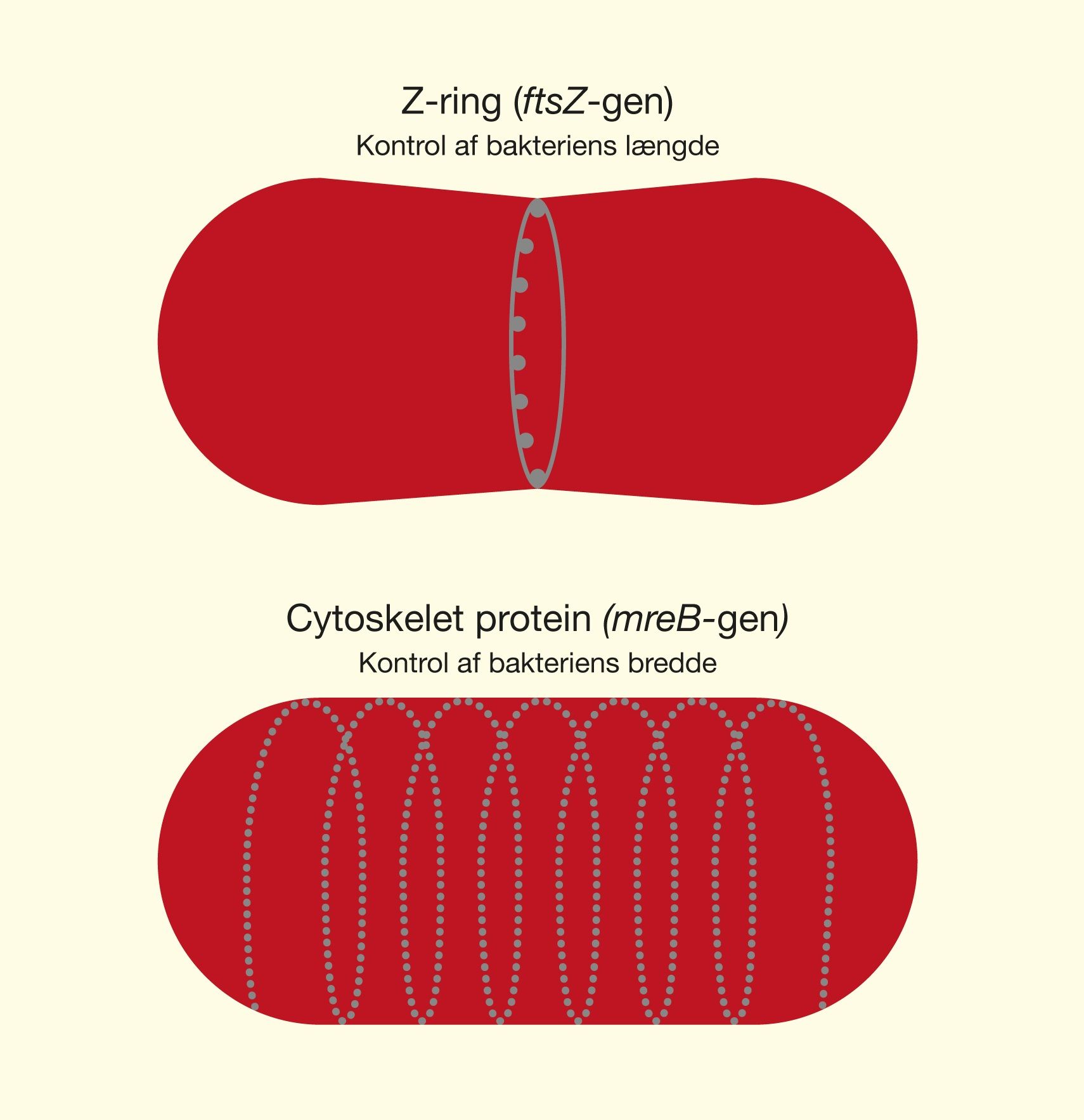
    The ftsZ gene encodes a protein that plays a key role in cell division, where new cells are formed. The protein is called FtsZ and, together with other proteins, forms a Z-ring, which contracts and divides a bacterium into two new ones. The amount of the protein determines how long the bacteria can stay before they are divided. By inhibiting gene expression and thus lowering the amount of FtsZ protein, cell division is inhibited, whereby cells grow longer before dividing. The function of the ftsZ gene can be seen in Figure 20.

    The mreB gene encodes the MreB protein, which has the function of regulating the size of the bacterial cytoskeleton, which determines the spatial structure of the cell. By inhibiting the expression of the mreB gene, the production of the MreB protein is slowed down, which means that the size cannot be regulated normally. This means that the cell can grow larger, although with a more weakened cytoskeleton. The function of the mreB gene can be seen in Figure 20.

    Figure 20. Here you can see the function of the studied genes, ftsZ and mreB. By inhibiting the ftsZ gene, the Z-ring cannot form to the same degree and the cells will become longer. By inhibiting the mreB gene, the associated protein cannot regulate cell size to the same extent and thus the cells can become fuller.

    In order to perform this gene silencing with dCas9, sgRNA had to be designed that could identify the two genes so that dCas9 could bind to them. Here, the researchers selected 20 nucleotides that were complementary to the DNA sequences of the genes, just as it was reviewed in the section Design of sgRNA, under Basic Theory. Since the binding of dCas9 generally provides the best efficiency on the coding string, sgRNA was designed to be complementary to it. Multiplexing was used to make dCas9 hit multiple sites on the genes. The reason was to investigate which combinations of binding sites for dCas9 provided the most effective reduction of gene expression. For each gene, 5 different sgRNA were designed that could target different areas of the genes. A lot of different combinations of these different sgRNA were tested for each gene to find the most effective combination for each gene. This multiplexing is done by introducing one or more pieces of sgRNA together with the gene for dCas9 into a vector, similar to what was discussed in the section Delivery methods, under Basic Theory.

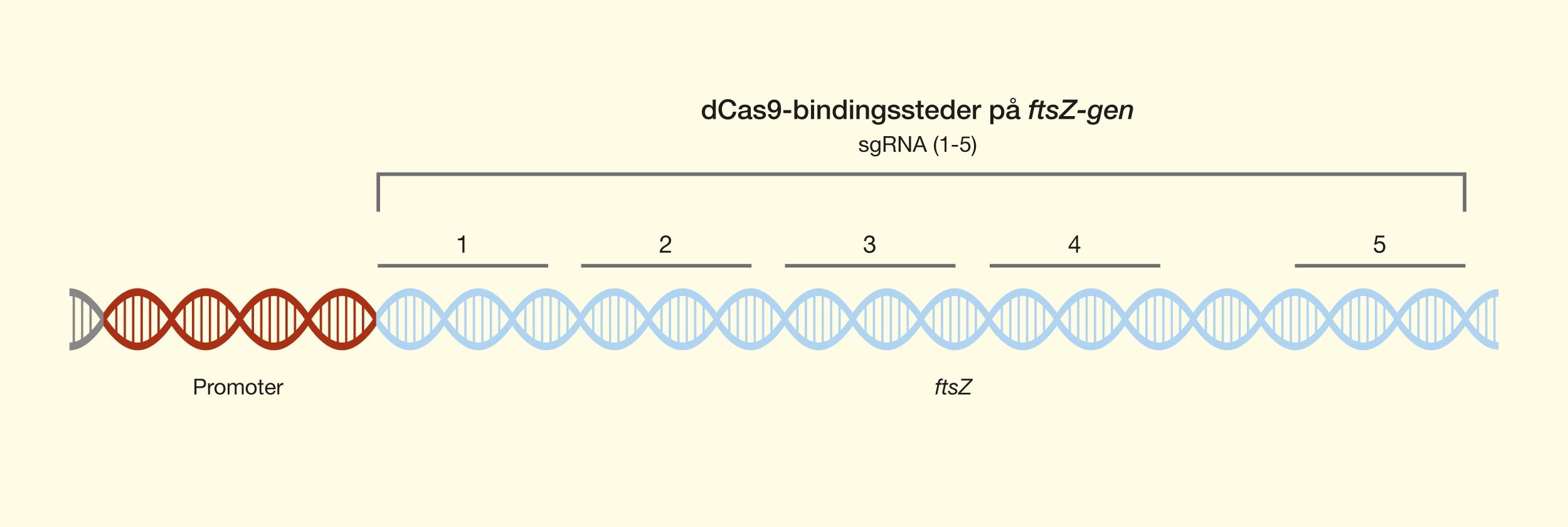
    For the ftsZ gene, sgRNA was designed to perform blocking of the transcription elongation, in which dCas9 binds in the middle of the gene and prevents the movement of RNA polymerase along the DNA strand. This is shown by the 5 dCas9 binding sites shown in Figure 21.

    Figure 21. The location dCas9 binding sites on the ftsZ gene for the 5 different sgRNA.

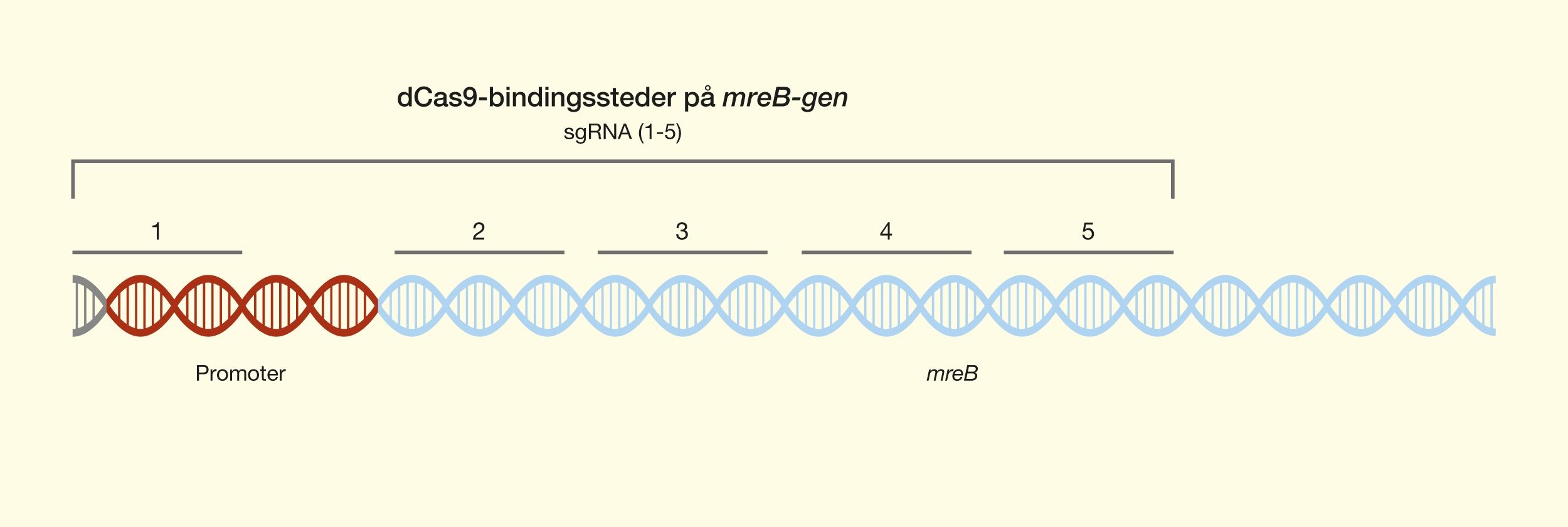
    For the mreB gene, sgRNA was also designed to block the transcription extension, as well as the blocking of the transcription initiation, where dCas9 binds in the promoter and prevents the binding of the RNA polymerase. This is shown in Figure 22, which shows the 5 dCas9 binding sites on the mreB gene.

    Figure 22. The location dCas9 binding sites on the mreB gene for the 5 different sgRNA.

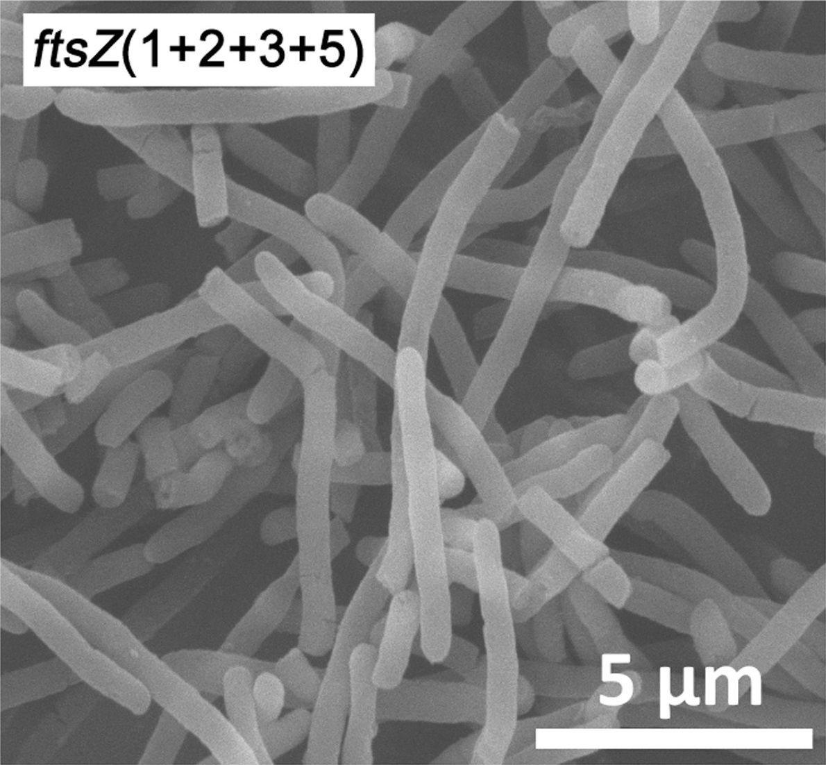
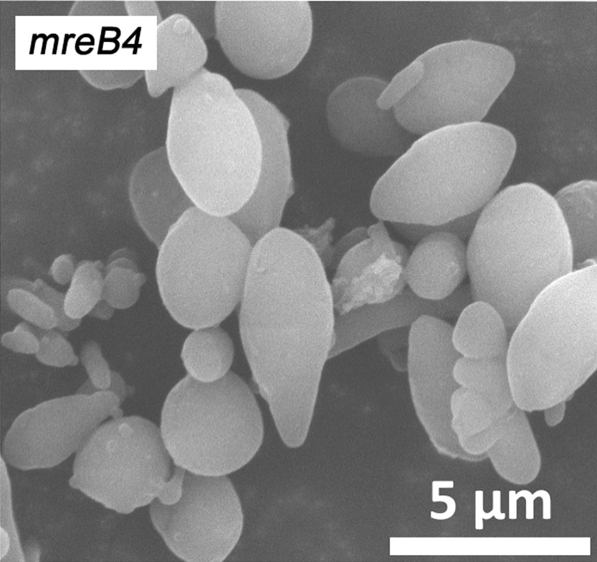
    Selected CRISPRi-modified E.coli bacteria are seen in Figure 23, for the ftsZ gene, and Figure 24, for the mreB gene, where it can be clearly seen that their morphology has changed due to the repression of gene expression. There simply isn’t enough mRNA transcribed to maintain the wild-type phenotype.

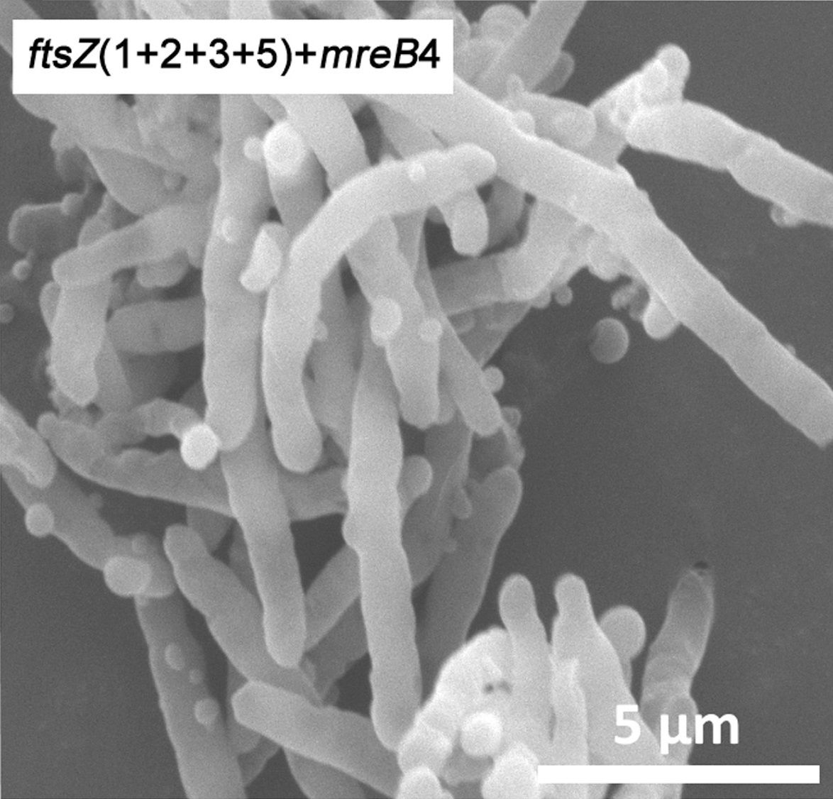
    For the ftsZ gene, multiplexed repression is observed, where 4 sgRNA has been used to cause binding of dCas9 at positions 1, 2, 3 and 5, as shown in Figure 21. It can be seen that this has led to significantly longer bacteria.

    For the mreB gene, a single piece of sgRNA was used to hit position 4, as indicated in Figure 22, causing swollen bacteria.

    In all these bacteria, dCas9 is bound to the selected positions in the DNA of the two genes, as can be seen in Figure 18. This leads to variations in morphology, because the genes have a reduced expression due to CRISPRi gene silencing.

    Figure 23. CRISPRi gene silencing of the ftsZ gene at dCas9 binding sites No. 1,2,3 and 5, as shown in Figure 21, produces altered morphology of E. coli. Longer bacteria are obtained by multiplexed reduction of the expression of the ftsZ gene.

    Source: “CRISPRi engineering E. coli for morphology diversification“, D. Elhadi, L. Lv, X.R. Jiang, H. Wu, G.Q. Chen, Metabolic Engineering, 38:358-369 (2016).

    Figure 24. CRISPRi gene silencing of the mreB gene at dCas9 binding site No. 4, as shown in Figure 22, produces altered morphology of E. coli. More full-bodied bacteria are obtained by reducing the expression of the mreB gene.

    Source: “CRISPRi engineering E. coli for morphology diversification“, D. Elhadi, L. Lv, X.R. Jiang, H. Wu, G.Q. Chen, Metabolic Engineering, 38:358-369 (2016).

    The researchers also tried to combine the repression of both genes, which is an example of multiplexing several different genes. Here they used 5 sgRNA at the same time, of which 4 caused dCas9 binding on the ftsZ gene and the last piece of sgRNA targeted the mreB gene. The result of this double gene silencing can be seen in Figure 25, where it is very clear that the E.coli bacteria do not have their normal morphology at all.

    This was an example of how CRISPRi can be used to perform gene silencing and thus reduce the expression of genes. As the images attest, binding dCas9 to specific sequences can produce drastic changes in the phenotype of an organism. These changes are achieved without destroying the DNA sequence, but simply by regulating how genes are expressed.

    Figure 25. CRISPRi gene silencing of the ftsZ gene at dCas9 binding sites No. 1,2,3 and 5, and the mreB gene at dCas9 binding site No. 4. Here we see a major change in the morphology of E. coli, as there is a reduction in the expression of both genes, ftsZ and mreB, which makes the bacteria both longer and fuller.

    Source: “CRISPRi engineering E. coli for morphology diversification“, D. Elhadi, L. Lv, X.R. Jiang, H. Wu, G.Q. Chen, Metabolic Engineering, 38:358-369 (2016).

    dCas9 with effector domains – repression (CRISPRi) and activation (CRISPRa)

    Until now, the use of dCas9, as a physical blockade to reduce the expression of genes, has been reviewed. In fact, dCas9 can be used for more than just a blockade that settles on DNA strands. It is possible to use dCas9 to manipulate the natural regulation of gene expression. Using this approach, it is actually also possible to upregulate the expression of genes, thereby increasing the amount of proteins that the genes encode. This type of upregulation is called CRISPR activation (CRISPRa).

    Eukaryotic genes contain small regulatory sequences that sit near the promoter. These regulatory sequences can either lead to upregulation of gene expression, whereby the gene is expressed more, or to downregulation of gene expression, whereby the gene is expressed less. There are sequences called enhancers that can upregulate gene expression and silencers that can downregulate gene expression. For regulatory sequences to have an effect on gene expression, certain proteins must bind to them, called transcription factors, that either turn the transcription up or down. If it binds to an enhancer and turns up the gene expression, it is called an activator. If, on the other hand, it binds to a silencer and turns down the gene expression, it is called a repressor. If you imagine gene expression as a moving car, you could say that activators act as the accelerator and repressors as the brake. It is these activators and repressors that are interesting to exploit together with dCas9.
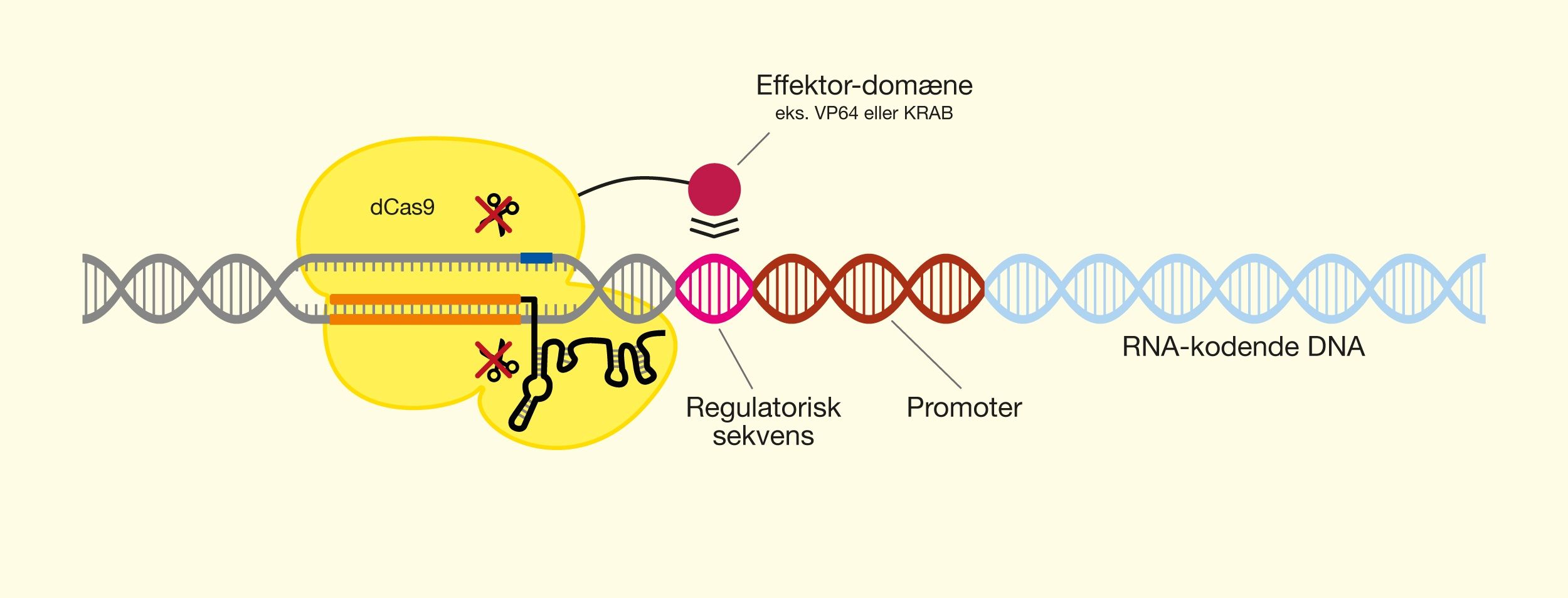
    You can use dCas9 as a supplier of transcription factors by attaching their active protein domains to the surface of the protein. These protein domains are generally referred to as effector domains. Next, you give dCas9 a piece of sgRNA that recognizes a place near a regulatory sequence where dCas9 can then bind. From here, the active effector domain can play its role as a regulator of gene expression, which can have an effective effect on the control of the gene. dCas9 thus becomes an sgRNA-controlled supplier of proteins that regulate gene expression. CRISPRi is executed by repressor effector domain while CRISPRa is executed by an activator effector domain. The structure of dCas9 with effector domains can be seen in Figure 26.

    Figure 26. dCas9 with merged effector domain to regulate gene expression. The regulatory sequence is influenced by the active domain and regulates transcription. If the domain is an activator, the transcription will be upregulated (CRISPRa), while a repressor will cause downregulation (CRISPRi).

    Below are two examples of dCas9 where effector domains have been merged that regulate the transcription of genes.


    Cas9-KRAB – downregulation of gene expression (CRISPRi)

    A Krüppel associated box (KRAB) is a protein domain found on many transcription factors that control human genes. Its purpose is to reduce gene expression and thus it acts as a repressor. By putting a KRAB protein domain on dCas9 and directing it towards genes’ regulatory sequences with sgRNA, you can choose which genes should have reduced gene expression. In this way, one can control the repression of specific selected genes, which has been done in human cells.


    dCas9-VP64 – upregulation of gene expression (CRISPRa)
    A VP64 protein domain is an artificially produced activator that can upregulate gene expression. When fused on dCas9, it can also be directed against specific genes’ regulatory sequences with sgRNA. This makes it possible to upregulate gene expression of selected genes. This has also been done in human cells.

    Examples of practical applications of CRISPRi and CRISPRa


    Genome screening and examination of gene functions

    Since dCas9 can be easily used and programmed with sgRNA, it is a powerful tool for searching for genes in whole genomes. sgRNA is easy to produce artificially, and thus sgRNA libraries can be created. These contain many different sgRNA, which can search for the genes you want to investigate. The function of the wanted genes can be investigated by utilizing CRISPRi, to reduce transcription, or CRISPRa, to upregulate the transcription and see how it affects the phenotype. In these methods, the DNA sequence is preserved, unlike genetic testing with Cas9 knock-outs or knock-ins.


    Control over the transcription of specific genes

    You can create types of dCas9 with effector domains that can be switched on and off, so you can determine when the gene should be repressed or activated. For example, a type of dCas9-VP64 system has been created that can activate transcription when the cells are illuminated with blue light. Each time the cells are exposed to blue light, dCas9-VP64 becomes active and upregulates the transcription of the gene. So there is stronger control over transcription.


    Artificial differentiation of cells

    Gene expression controls the differentiation of cells into more specialized cells, such as when a stem cell develops into a subtype of cell. CRISPRi and CRISPRa can be used to control gene expression, thereby influencing the identity and differentiation of the cell. This controlled cell-type reprogramming has been used to transform connective tissue cells from mouse embryos into skeletal muscle cells. One of the main goals of this CRISPR technology is to be able to form stem cells that can be used for disease models or even for the therapy of diseases.

  • Case 3 - Gene therapy with CRISPR/Cas9

    Gene therapy – therapeutic gene editing with CRISPR/Cas9

    This section will discuss how CRISPR can potentially be used as gene therapy for genetic diseases in the future. Some concrete examples of genetic diseases and their potential treatment will be reviewed.


    Genetic diseases

    Genetic diseases occur due to errors in the genome. These errors are mutations in the DNA that may have been inherited or occurred during one’s life. The causes of genetic diseases can be found at very different levels of size. It can be changes in anything from whole chromosomes to single base pairs, but be equally serious. One can categorize genetic diseases according to their extent of cause.

    Monogenic diseases are caused by mutations in individual genes.

    Polygenic diseases are due to interactions between several genes.

    Chromosomal diseases arise from structural defects in the chromosomes or from abnormal numbers of the chromosomes.

    The phenotypes of genetic diseases are caused by the influence of mutations on genes. The influence can either be a change in the structure of the gene product (RNA or protein) with which it works incorrectly, or be changes in the amount of gene product produced at which the dose deviates from normal.

    One could, for example, imagine a monogenic disease in a hypothetical gene X that produces the protein X, which normally transports large necessary molecules into our cells. A mutation in gene x that causes a change in the structure of protein X could mean that protein X could not bind to the necessary molecules, with which nothing would be transported into cells. Another mutation in gene x, which led to an overproduction of well-functioning protein X, could mean that too many of the necessary molecules would be transported into the cells and cause a toxic effect. In both cases, one could imagine that the mutations would trigger a disease phenotype.


    Gene therapy

    Gene therapy is the modification of a patient’s disease-causing DNA, or RNA, for the purpose of preventing, curing or treating a genetic disease. The idea of gene therapy is that the correction of mutated genes will restore the original function and eliminate the genetic disease. Genetic modification can, for example, take place by destruction, insertion, excision or replacement of the disease gene, depending on the culprit mutation, as described in the basic theory section Molecular strategies. The point is that you want to correct the genotype to eliminate the disease phenotype in question.
    Monogenic diseases are usually more tangible for gene therapy purposes, as they are only caused by mutations in single genes. The gene therapy has therefore had a strong focus on the monogenic diseases and gene correction of these with CRISPR/Cas9 is therefore most well researched. Although CRISPR technology has the potential to revolutionize gene therapy and that the development of the technology is happening at a rapid pace, there is still some way to go. There are some challenges that need to be overcome and more research is required before CRISPR/Cas9 becomes practical for gene therapy.

    There are two main approaches to performing a gene therapy correction of disease genes with Cas9. In vivo means “inside the living”, and refers to the correction taking place inside the living organism itself. Ex vivo means “outside the living”, which refers to the correction taking place outside the organism, i.e. in cells that have been removed from the organism itself.


    In vivo gene correction

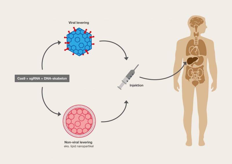
    The gene therapy takes place in the patient by direct delivery of the gene correction system: Cas9, sgRNA and possibly a DNA template. The transfer of the system to the patient can take place by different methods. This can be done by local injection, meaning that the therapy takes place in a specific organ or defined tissue. A systemic injection causes the components to enter the bloodstream and will spread throughout the body. As soon as the components are transferred to the cells, the gene editing will take place. This transfer of the components into the cells themselves can take place in different ways, for example with a viral vector or lipid nanoparticles. See Figure 27 for in vivo gene correction. A safe, efficient and controlled delivery of the system is important to ensure that the gene correction takes place correctly and in the correct tissue. This is very important aspect and one of the major challenges for gene therapy with CRISPR/Cas9. The different forms of delivery (DNA-, RNA- and protein-based) are described in the basic theory section Delivery methods.

    4.1

    Figure 27. In vivo gene correction. CRISPR/Cas9 is delivered directly into the patient’s tissue, with which the gene correction takes place inside the patient. The transfer can take place in different ways, for example through viral delivery or lipid nanoparticles, and with different components, such as a DNA template or several pieces of sgRNA.

    The gene correction can be done at different cell levels, giving different results and consequences. The correction can occur in the zygote (the fertilized egg cell) and in the early embryonic stages, at which point all daughter cells formed by subsequent cell division will inherit the correction of the gene. This may ultimately mean that all cells in the organism will contain the gene correction, including the gametes, which means that the gene correction is hereditary. The correction of the gene can also take place in the somatic cells of the developed organism, with which only the affected cells and their daughter cells will have the correction. These gene corrections will therefore only be found in the affected tissue and will not be hereditary. Genetic modification contributes with very potent methods to eliminate genetically inherited diseases, but ethical issues make it a sensitive area of research.


    Ex vivo gene correction

    The gene therapy takes place in cells that have been extracted from the patient. The genetically modified cells with the desired gene correction can be selected and cultured outside the body, after which they are then transplanted back to the patient in the affected area. The cells with the gene correction will then perform the normal functions that cells with the disease gene previously could not. There are two main ways to perform an ex vivo gene correction, which depend on the types of the patient’s cells used as a starting point.

    Stem cells
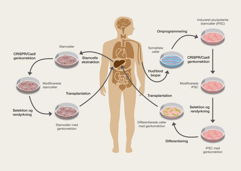
    Stem cells are first extracted from the patient himself. The gene correction is then made with CRISPR/Cas9. The genetically modified stem cells can then be transplanted back into the affected area of the patient’s body, where they form new differentiated cells. The newly formed specialized cells will contain the gene correction, as they originate from the corrected stem cells and will therefore be able to restore normal functions and treat the genetic disease. The stem cells themselves can also be differentiated into specialized cells, and then be transplanted back as differentiated cells in the affected area of the patient. See Figure 28.

    Somatic cells
    The cells used are fully developed somatic cells, such as skin cells or blood cells. The cells can be extracted and then genetically modified with CRISPR/Cas9, after which they are transplanted back to the disease area. However, these cells do not have to be from the area where you are interested in performing the gene correction, as they can be reprogrammed into other cell types. Somatic cells can be easily obtained from the patient by a blood test or biopsy of the skin. Subsequently, the cells are reprogrammed into induced pluripotent stem cells (iPSC), which are artificially generated stem cells. These iPSCs are genetically modified outside the body with CRISPR/Cas9 so that the disease gene is corrected. The corrected iPSC is isolated and differentiated into the type of cells found in the affected area. After this, the modified cells are transplanted back into the patient. See Figure 28.

    4.2

    Figure 28. Ex vivo gene correction. On the left are stem cells extracted from the patient’s tissue. These are modified with CRISPR/Cas9, after which the desired cells are selected and cultured. Eventually, the cells are transplanted back into the patient’s tissues. To the right is a biopsy of skin or blood cells differentiated into induced pluripotent stem cells (iPSC). These are modified, after which cells that have been modified correctly are selected and cultured. The iPSC is then differentiated into the cells of the given tissue type and these are transplanted back.

    A major advantage of ex vivo gene therapy is the ability to examine the gene correction in the cells outside the body before they are transplanted back. By selecting, you can select cells with correct modifications for the transplant and clean these, as well as throw away the cells that have been wrongly mutated. This quality check lowers the risk of side effects due to unintended mutations, such as Cas9 off-target effects, which cannot be easily detected by in vivo gene correction. A disadvantage of ex vivo gene correction is the requirement to be able to grow the extracted cells outside the body, which can make the process more complicated.

    Retinitis Pigmentosa

    Retinitis pigmentosa is a monogenic disease that causes progressive distortion and loss of vision. The symptoms are due to the degeneration of the eye’s photoreceptors (cone cells and rod cells). A typical disease course starts with night blindness, which develops into loss of mid-peripheral vision in early adulthood and ends with very limited tunnel vision that can make the sufferer legally blind. Other symptoms can also include incorrect colour vision and loss of visual acuity. The symptoms and their severity can vary greatly among those affected.
    At least 45 genes may contain mutations that cause the disease. The retinitis pigmentosa GTPase regulator (RPGR) gene encodes a protein that is related to the cilia of photoreceptors and is necessary for normal vision. Mutations in RPGR can cause a powerful version of Retinitis pigmentosa, which has an X-linked inheritance and therefore hits men hardest. A version of RPGR, expressed mainly in retinal photoreceptors, contains an exon called ORF15. Since exon ORF15 contains a mutational hotspot, mutations therein are typical. In a genetic study, 58 families were studied, all of whom were affected by retinitis pigmentosa. It was found that the exon ORF15 hotspot is approximately 500 bp long and that mutations in it were both deletions (max. 36 bp) and insertions (max. 75 bp). The mutations result in a dysfunctional protein, which causes the degeneration of the photoreceptors.


    Potential ex vivo gene therapy

    You can correct these mutations with CRISPR gene therapy by modifying the mutated sequence back to the wild-type sequence. A research team extracted skin connective tissue cells from a retinitis pigmentosa patient and converted them into induced pluripotent stem cells (iPSC), which were corrected with CRISPR gene therapy. These iPSCs could theoretically become cells that could be transplanted back into the patient and alleviate the disease, making this intervention a potential ex vivo gene therapy.

    In the patient’s ORF15-exon, there was a point mutation, TAG, that encodes a stop codon and thus causes a premature inappropriate stop of translation. The protein could therefore not be fully produced. The gene correction consisted of converting the point mutation into the wild-type sequence, GAG, which codes for the amino acid glutamate.
    DNA-based delivery was utilized to get the gene correction system into cells. Cas9 and sgRNA were delivered in a plasmid, while a DNA template was delivered in the form of a single DNA strand. When using homology directed repair, the mutated sequence was replaced with the DNA template. This strategy worked in 13% of the sequences in the treated cells. With ex vivo gene correction, one could imagine that a properly modified cell would then have been isolated, safety checked for potentially dangerous off-target effects and cultured into a larger amount of differentiated cells that could eventually be transplanted into the patient’s eye.

    Duchenne muscular dystrophy

    Duchenne muscular dystrophy is a monogenic disease caused by mutations in the gene DMD that normally produces the protein dystrophin. The disease has an X-linked recessive inheritance, which means that men will be severely affected by the disease and that women will generally be carriers of the disease, or have milder symptoms. About 1 in 5000 men get this disease. This disease causes a serious progressive weakening and breakdown of the body’s muscles (skeletal, cardiac and smooth). The course of the disease starts with mild motor problems, which develop into major movement problems and usually end with either the heart muscle or the respiratory muscles stopping functioning. The average life expectancy is 26 years.

    Dystrophin is an elongated structural protein in muscle tissue that, in collaboration with other proteins, has the function of connecting the muscle fibers and protecting them from damage when contracted and extracted. The protein consists of a long stabilizing domain that at both ends has domains that can anchor themselves to other proteins. Put simply, dystrophin acts as a shock-absorbing structural element. Complete lack of functional dystrophin causes Duchenne’s muscular dystrophy, while minor structural defects of dystrophin provide the relatively milder version of the disease, called Becker’s muscular dystrophy.

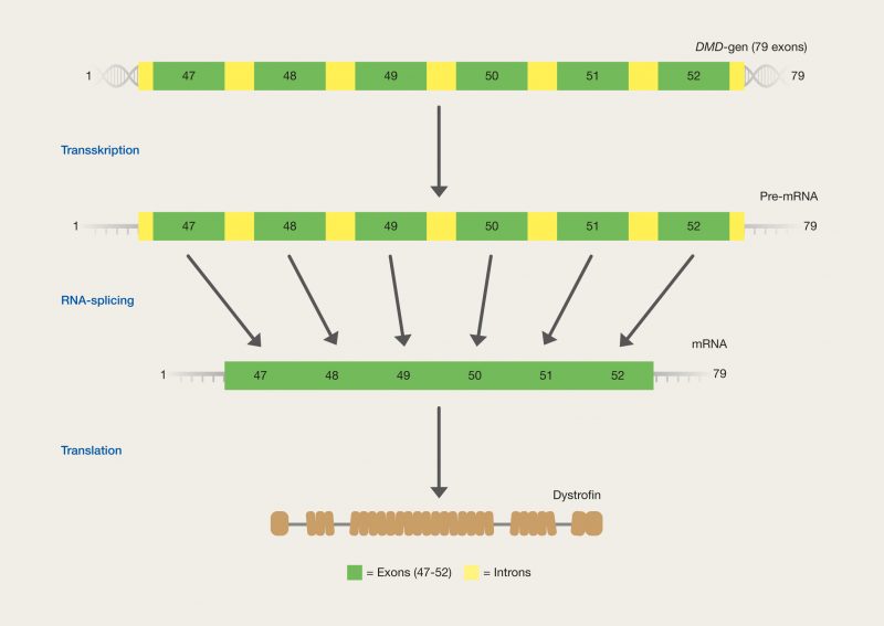
    The DMD gene is the largest known gene in humans (2.6 million base pairs) and is made up of 79 exons. Exons are sequences that contain code for the finished mRNA, while introns don’t encode anything. After transcription of the gene, all introns are excised from pre-mRNA, after which all exons are composed by a process called RNA splicing. The total exons make up the coding mRNA, which when translated forms dystrophin. The normal transcription, RNA splicing and translation of the DMD gene (exon 47-52) into functional dystrophin can be seen in Figure 29.

    4.3

    Figure 29. The normal function of the DMD gene. Here is a sample of the gene, from exon 47 to exon 52. Transcription forms pre-mRNA, and the formation of mRNA from pre-mRNA occurs by RNA splicing, where introns are removed and exons are assembled. mRNA is eventually translated into the functional protein dystrophin, which is essential for the normal functioning of muscles.

    Severe mutations in the DMD gene cause Duchenne muscular dystrophy. In approximately 60% of cases, the mutations in DMD are major deletions that cause frame-shift. This produces completely dysfunctional protein, which causes the violent phenotype. In the rest of the cases, it is other mutations that also cause frame-shift or sit in the important domains of the protein and make it completely dysfunctional in that way. More than 3000 different disease-causing mutations have been identified for the disease and many of these are located in hotspots, i.e. specific exons that are particularly vulnerable.

    Becker’s muscular dystrophy is linked to in-frame deletions in the long dystrophin domain. Here, the reading frame is not destroyed, since base pairs in a multiple of 3 nucleotides are removed. Dystrophin is therefore only shortened and retains a slightly reduced functionality, which means that the disease phenotype becomes relatively mild.

    The genetic background to Duchenne muscular dystrophy is complex and there is no single genetic intervention that can cure the disease. Therefore, different CRISPR-based strategies have been tested on the different types of mutations. An insight will now be taken into two strategies, both based on removing a segment of the gene that causes the disease. It is known from Becker’s muscular dystrophy that in-frame deletions produce milder symptoms than frame-shift deletions that cause Duchenne muscular dystrophy. The idea is to remove the exons that contain the frame-shift mutations with Cas9, which will artificially create an in-frame deletion of the given exon. The bad Duchenne’s muscular dystrophy is thus transformed into the relatively milder Becker’s muscular dystrophy.


    Ex vivo gene correction: removal of frame-shift mutations

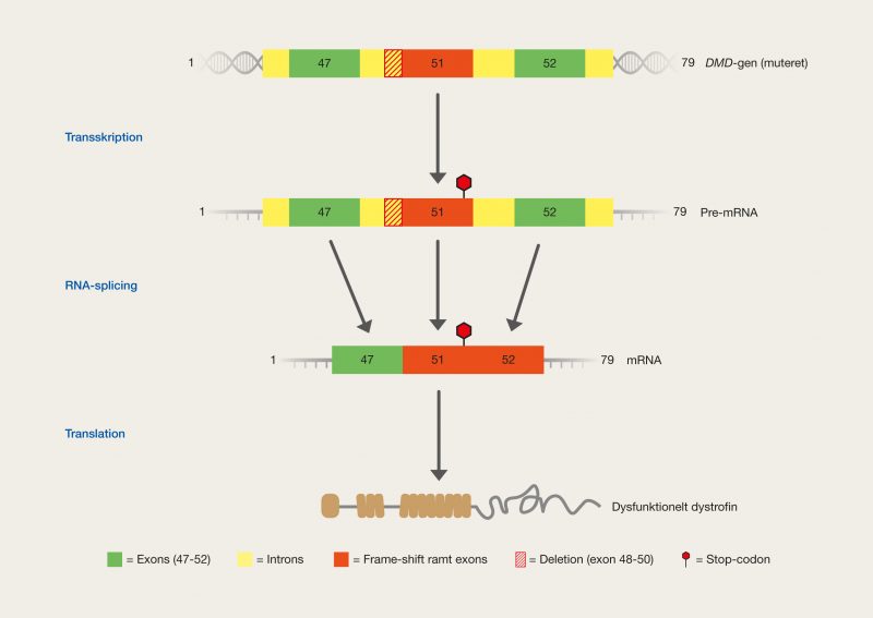
    The larger deletions that cause frame-shift mutations are often located in specific hotspot exons of the DMD gene. The important thing for the correction of such an exon is to prevent the frame-shift mutation from completely destroying the protein and instead achieve the production of a functional alternative dystrophin. A patient affected by Duchenne muscular dystrophy has a deletion of hotspot exons 48 to 50 that causes a frame-shift mutation in exon 51, forming a misplaced stop-codon. Transcription, RNA splicing and translation of the mutated DMD gene (exon 47-52) into dysfunctional dystrophin can be seen in Figure 30. Note that the formation of a stop codon is caused by a displaced reading frame, due to the frame-shift mutation that is a consequence of the deletion. This means that the translation of dystrophin cannot be done correctly.

    4.4

    Figure 30. It mutated the DMD gene in a patient with Duchenne muscular dystrophy. Here is a sample of the gene, from exon 47 to exon 52. The frame-shift mutation that has occurred in exon 51, due to a deletion of exon 48 to exon 50, causes a stop-codon. Transcription forms pre-mRNA, which contains a stop codon, and the formation of mRNA from pre-mRNA occurs by RNA splicing, where introns are removed and exons are assembled. The finished mRNA still contains a stop codon and translation is therefore stopped prematurely, leading to dysfunctional dystrophin, which causes Duchenne muscular dystrophy.

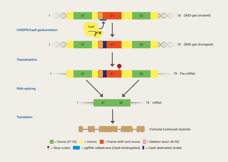
    By mutating the RNA splice site between the mutated exon and the preceding intron, one can skip the use of the mutated exon in the finished mRNA, as RNA splicing cannot take place normally. Thus, the consequence of the mutation, i.e. the stop codon, will not be included in the finished mRNA. The mRNA produced will then encode a shorter, but functioning, version of dystrophin, thereby making the disease phenotype milder. This strategy is called exon skipping because we actively opt out of the use of specific exons. This is done by destroying the sequence, by exploiting Cas9 and NHEJ.
    sgRNA was designed to hit the boundary between exon 51 and the preceding intron, with which Cas9 introduced an indel mutation into the RNA splice site. This caused the mutated exon 51 to be ignored by RNA splicing and the new mRNA to have an in-frame deletion of exons 48-51. This allowed functional dystrophin to be produced, albeit with a reduced version missing the parts encoded by the Exon 48-51. Gene correction, as well as transcription, RNA splicing and translation of the corrected DMD gene (exon 47-52) for functional dystrophin can be seen in Figure 31.

    4.5

    Figure 31. DMD gene in a patient with Duchenne muscular dystrophy, and gene correction (exon-skipping) with CRISPR/Cas9. Here is a sample of the gene, from exon 47 to exon 52. The frame-shift mutation that has occurred in exon 51 due to a deletion of exon 48 to exon 50 causes a stop codon. Cas9 is used to destroy the RNA splice site that handles exon 51. Transcription forms pre-mRNA, which contains a stop codon. The formation of mRNA from pre-mRNA occurs by RNA splicing, but the mutated exon 51 is not included in the collection of exons, as the Cas9-induced mutation means that RNA splicing ignores it. The finished mRNA does not contain a stop codon as exon 51 has been removed by exon skipping and translation therefore functions normally, resulting in the production of a truncated, but functional, version of dystrophin.

    With this strategy, we succeeded in restoring the production of dystrophin in iPSCs from human heart muscle cells, thereby restoring the contractor function of the heart muscle. This gene modification has great potential for cases of Duchenne muscular dystrophy, which is due to frame-shift deletions, by correcting muscle cells to cause milder symptoms. This could be the basis for a potential ex vivo gene therapy.


    In vivo gene therapy in mouse models: excision

    Diseases can be recreated as models in other organisms, such as mice. The mdx mouse model of Duchenne muscular dystrophy contains a mutation in exon 23 of the dystrophin gene that produces an inappropriate stop codon. The dystrophin protein cannot therefore be fully translated.

    Removing the mutated exon 23 means that the protein can be translated into a shortened and functional version of dystrophin, just as seen in the previous example. Cas9 has been used to perform such an excision of the whole exon 23, by multiplexing by two sgRNA, after which the NHEJ can compose the gene. The two pieces of sgRNA were designed to hit in the intron sequences on either side of exon 23.
    The gene correction system was delivered by DNA-based delivery through viral vectors, which were introduced by both local and systemic injection into the mouse models. The gene correction is therefore in vivo. The virus used was adeno-associated virus, which can only contain a limited amount of DNA with which the system might be divided. One quantity of virus was packed with Cas9, while another quantity of virus was packed with the two pieces of sgRNA.
    Using this strategy, dystrophin production could be restored in the mouse models, thereby improving the structure and function of the muscles. A few unexpected side effects were found, but by further optimizing the effectiveness and safety of the system, Cas9 has great potential as gene therapy in this type of genetic disease.

    CRISPR technology in cancer research

    Cancers are critical and can cause many unpleasant symptoms, as well as have serious consequences if not fought. Cancers are called multifactorial, as they can be caused by the interaction of many different factors, both genetic and environmental. These causes can vary greatly between individual cases and therefore cancer is a very complex disease to fight.
    The use of CRISPR technology is interesting in cancer research, due to the many different genetic conditions of cancer, which can be investigated and hopefully treated with this new promising technology in the future.

    Some main aspects of cancer genetics are oncogenes and tumor suppressor genes, which are single genes that have a direct effect on the development of a cancer.

    Oncogenes accelerate the development of a cancer disease. The gene may have become an oncogene because it becomes overexpressed, thus making too much protein, or because a gain-of-function mutation has emerged that gives the resulting protein a new carcinogenic property. In both cases, cancer develops.

    Tumor suppressor genes usually suppress cancer, with which the lack of the genes causes cancer. Tumor suppressor genes may lose their effectiveness because they are underexpressed, thus making too little protein, or because a loss-of-function mutation has occurred that removes the cancer-suppressing property from the protein.

    Metaphorically, you could say that oncogenes are accelerators for cancer that is pressed harder and tumor suppressor genes are brakes for cancer that is released.


    Ovarian cancer

    Ovarian cancer can occur in women’s ovaries, where both egg cells and female sex hormones are produced. In Denmark, ovarian cancer affects 553 new women every year. Ovarian cancer can occur on the basis of many different factors, but here we will take a look at the DNMT1 gene. DNA methyltransferase 1 (DNMT1) is an important enzyme for the epigenetic maintenance of DNA. The normal function of the enzyme is to put methyl groups on DNA (methylation). It has been shown that overexpression of DNMT1 leads to inhibition of tumour suppressor genes, which cannot control cell growth. Uncontrolled growth of cells is a hallmark of cancer.

    Destruction of the DNMT1 gene in cancer cells can stop the inhibition of tumour suppressor genes so that they can actively retain the cancer. In vivo gene therapy for this type of ovarian cancer has been tested in mouse models specifically designed for disease modelling of cancer. The experimental mice have a type of mutation that means that they cannot develop the sprat, which is an important organ for the immune system. This allows the mice to be easily transplanted with promoted tissues, such as human cancer cells, making them very important laboratory animals for cancer research. The experimental mice were transplanted with human ovarian cancer cells.
    The DNMT1 gene is important for the normal function of cells, and therefore it is not desired that the gene be destroyed in healthy cells. In vivo gene therapy must therefore target cancer cells. This is achieved by making sure that the gene correction system is only delivered to the cancer cells. DNA-based delivery was used, where a plasmid coding for Cas9 and sgRNA was packaged into nanoparticles, called liposomes. These liposomes were designed to search for the cancer cells specifically. Cas9 was used to destroy the DNMT1 gene in cancer cells, by designing a piece of sgRNA that specifically recognized the DNMT1 gene. Indel mutations, formed by destruction, made sure to inactivate the gene.
    This strategy could successfully inhibit the spread of cancer and kill some of the cancer cells. The approach has the potential as an in vivo gene therapy that can inhibit the development of ovarian cancer, but a great deal of work is required to streamline the correction so that dangerous side effects are avoided.

    Complications and challenges of gene therapy with CRISPR technologies

    Complications that exist with the CRISPR/Cas9 technology itself are obvious problems, such as inefficiencies of homology directed repair due to NHEJ. One of the biggest problems is off-target effects, as unexpected mutations can occur and cause bad side effects due to the lack of specificity. There are further complications associated with the use of CRISPR technology for gene therapy, which will be briefly elucidated here.


    In vivo versus ex vivo

    The ex vivo approach is more complicated and demanding than In vivo, due to maintenance, culture and return of the extracted cells, but In vivo is linked to several ethical problems due to possibilities for inheritance of the gene corrections. In addition, the effectiveness of in vivo genetic modification is significantly lower than that of ex vivo genetic modification, which also depends on the efficiency of delivery. Ex vivo gene therapy offers more opportunity for verification of genetic modification and there are more possibilities for combination with other technologies, as the In vivo approach is limited by taking place inside the body. More research into gene therapy is needed before these approaches become practical.


    Individual treatment requires individual gene therapy design

    The same genetic diseases can be caused by many different mutations, which means that they must also be treated with different gene modifications. This means that each patient must have specifically designed their own gene therapy. This can be a major challenge for the production of the gene therapeutic components, as many considerations must be taken into account. For example, each sgRNA must be designed after each mutation to be corrected, subject to minimal off-target effects, as well as maximum specificity and effectiveness.


    Precision and efficiency of delivery

    The packaging of all the components of a gene correction system to be used for gene therapy is a problem for both DNA, RNA and protein-based delivery. In DNA-based delivery using viral vectors, you may have to split the system into several virus particles. A challenge with protein-based delivery is the packaging of the large Cas9 protein together with both sgRNA and a possible DNA template. In vivo gene therapy requires greater control over where the gene correction system ends up, both by systemic and local injection. Another aspect is the amount of the dose of Cas9 and sgRNA needed for the disease to be cured.


    Immunological response to CRISPR/Cas9

    An important point for the use of CRISPR technology in humans is that Cas9 originates from a bacterium with which the immune system can be expected to react. Foreign proteins must be monitored by the immune system, as they can be potentially dangerous. The Cas9 protein can be eliminated by the immune system, but furthermore, the very human cells that express the Cas9 protein can be killed. With the use of DNA- and RNA-based delivery, a greater immunological response is expected than with protein-based delivery. This is due to the active expression of the alien Cas9 in the cells, from the introduced DNA or RNA, which can be longer-lasting. The presence of Cas9 is more short-lived when using protein-based delivery, as no more Cas9 is produced than has been introduced. The short-term presence provides less opportunity for an immunological response. In addition, introduced Cas9-coding DNA can integrate permanently into the DNA of cells. When using viral vectors, an immunological response can be expected in particular due to the pathogenic nature of the virus particles.

  • Case 4 - Gene drives to gene modification of populations

    Gene drives and automatic genetic modification of entire populations

    CRISPR technology allows the formation of an incredibly powerful genetic tool that can genetically modify entire populations by a chain reaction of mutations. This genetic tool is called a gene drive and utilizes CRISPR/Cas9 gene modification to manipulate the genetics behind gene inheritance.

    Genetics

    In eukaryotic genetics, diploid organisms are most often seen, which means that they have two copies of each chromosome type. These interrelated copies are called homologous chromosomes. Each copy comes from each of the parents, by which they are likely to be different. Since there are two copies of each chromosome, there are also two copies of each gene. These two copies are called alleles, and may be different, encoding different properties of the same gene. If two identical alleles are had, the organism is homozygous, but if two different alleles are had, the organism is heterozygous. The expression of alleles can be described as recessive or dominant. If the organism is heterozygous, dominant alleles will be expressed in the phenotype, while recessive ones will not. If the organism is homozygous, the alleles are the same and the shared property is expressed. The location of the gene on the chromosome, and thus the alleles, is called the locus.

    An example of this type of genetics could be blood types. There is one gene that codes for the blood type. On each homologous chromosome sits an allele of this gene. There are 3 different alleles: A (dominant), B (dominant) and 0 (recessive). You can have one allele on each of the 2 homologous chromosomes. The different alleles are located in the ABO locus of chromosomes. Let’s look at 3 examples of combinations:

    One allele codes for A blood group (dominant), while the other encodes 0-blood group (recessive), by which the person expresses blood group A. These each code for its own blood type, which is heterozygous, but blood group A is dominant and expressed.

    Both alleles encode the 0 blood type (recessive), by which the person expresses blood type 0. This is only possible because it is a homozygous with recessive alleles. There is no dominant allele present that can dominate over the recessive expression.

    If you have an allele for A blood group (dominant) and an allele for B blood group (dominant), you have a special situation called codominance. Here, the person expresses blood type AB, i.e. both A and B.

    Inheritance

    Genes are dynamic and are exchanged between organisms in several ways. Usually, genes are inherited according to the Mendelian laws of inheritance, which describe the further distribution of genes between generations. The laws of distribution state that the genes are inherited randomly, with equal probability to each offspring and independently of each other.
    Mendel’s 1st law – the law of division: In the formation of offspring, the alleles are separated from each other, and the offspring inherit one allele from the father and one allele from the mother. Each of the two inherited alleles can be each of the parents’ two alleles, with equal probability. Thus, each of two related alleles is distributed with a 50% probability to offspring.

    Mendel’s 2nd law – udependent selection: Alleles of different genes on different chromosomes are distributed independently.

    Of course, as with most other rules, there are exceptions, including nuisance drives.

    Gene drives

    Gene drives are a showdown against the Mendelian inheritance of genes, as these aim to promote the inheritance of specific alleles. You could say that it is a forced inheritance of specific alleles to all offspring, which is called super-Mendelian inheritance.
    Gene drives are selfish alleles that seek certain sequences into which they can copy themselves, destroying the original sequence. They spread themselves by taking advantage of the functions of the organism. Further, gene drives can carry on self-selected alleles that code for a given new trait or another genetic modification. These alleles do not necessarily have to be beneficial to the organism. In this way, gene drives can implement genetic changes in entire populations, by forced spread through the natural inheritance of genes over generations.

    Think of the normal Mendelian inheritance of genes as a coin toss, where there is a 50% probability of either heads or tails. Similar to a coin toss, one of each of the parents’ two alleles is randomly inherited. Gene drives are similar to a coin that always gives you the side of the coin you want. Gene drives remove the randomness of whether it will be one or the other allele and determine that it will always be the designed gene drive allele that is forced through. Thus, there is theoretically a 100% probability of inheritance, rather than 50%.

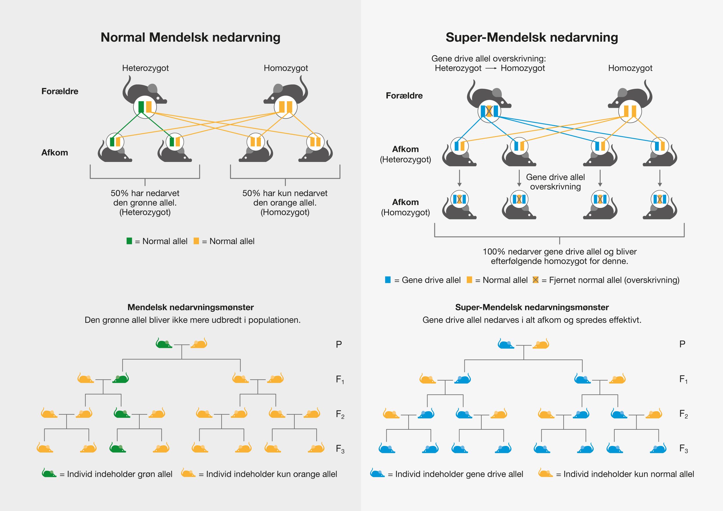
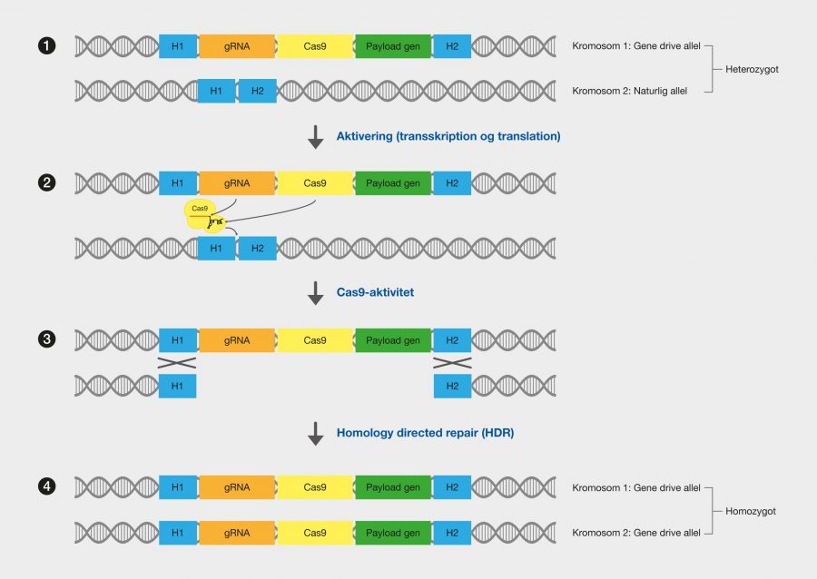
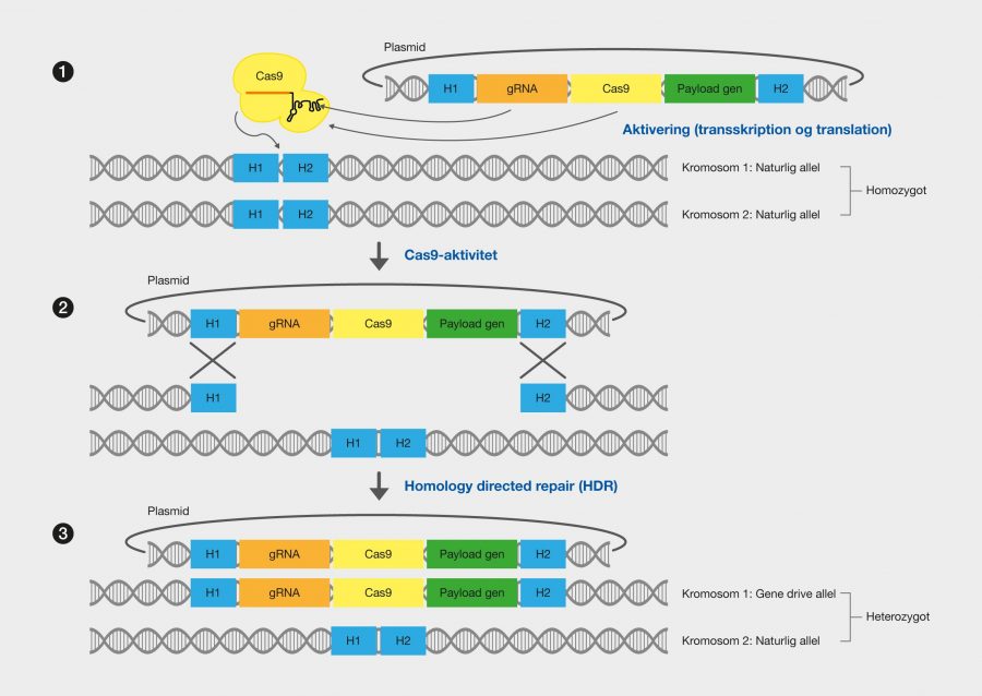
    This is done by gene drive alleles in a heterozygous individual copying themselves onto the other allele, thus creating an individual that is homozygous for the gene drive allele. This happens to all individuals who inherit the allele. Figure 32 shows how a gene drive takes the natural Mendelian inheritance by surprise by overwriting the natural alleles and creating homozygotes, which always pass on the gene drive allen, and how this manipulates the inheritance pattern to ensure gene drive allele inheritance.

    Figure 32. Normal Mendelian inheritance compared to super-Mendelian inheritance, as seen in the use of gene drives.

    Gene drives can be used to make gene knock-outs and knock-ins of self-selected genes on entire populations. This mutagenic process has previously been called Mutagenic Chain Reaction (MCR) due to its automatic and self-sustaining formation of mutations that spread as a chain reaction throughout the population. Based on the impact of genetic modifications on the population, gene drives can be divided into two types:

    Modification drives: designed with the purpose of modifying a population, for example by spreading specific genes among the population. These genes can give the organisms new characteristics, both beneficial and unfavorable.

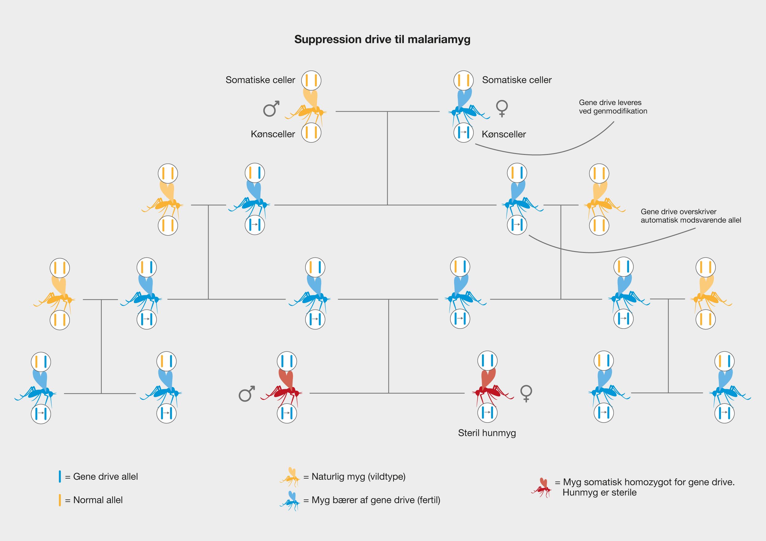
    Suppression is driven: designed for the simple purpose of reducing or completely eliminating a population, for example by destroying essential genes or producing infertility.

    Uses

    Gene drives are particularly useful in health, environment and agriculture. Many different species can have negative impacts on the environment and humans, such as by causing diseases, destroying crops or threatening other indigenous species. Therefore, it is an attractive idea to be able to control the fate of entire populations by self-sustaining genetic manipulation. Gene drives are designed for the specific situation with different genetic components so that they function optimally for the given purpose. Genetic engineering is incredibly efficient and powerful, which includes great responsibility. An important consideration is that once a gene drive has been released into a population, you cannot return to the completely natural genotype again if the gene drive is effective and implemented throughout the population. However, new gene drives can be spread that aim to restore the former phenotype.

    Prevent vector-borne diseases

    These diseases are spread by transmission from one organism to another. One can stop these diseases by, for example, eliminating the organism that causes the infection to humans, with suppression drives, or preventing the organism from containing the disease with modification drives. Examples of vectors for some known human diseases are:

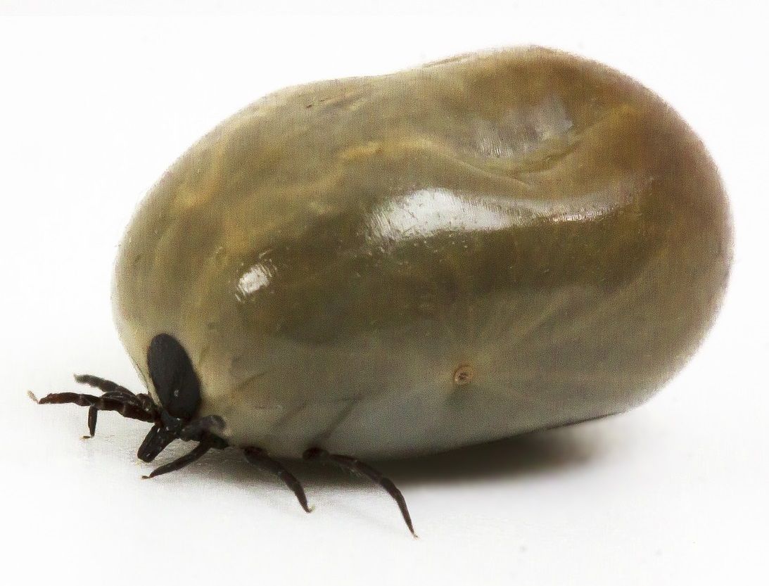
    Ticks (e.g. Lyme disease and tick-borne encephalitis).

    Lice (e.g. trench fever and typhoid).

    Fleas (e.g. plague and typhoid).

    Mosquitoes (e.g. malaria, dengue, yellow fever, West Nile virus infection and Zika virus infection).


    Eradicate invasive species or pests

    Alien species can significantly affect ecosystems by putting them out of balance, for example by outcompeting other native species. Suppression drives are smart here to eliminate the harmful population.
    Several risks are run by the use of suppression drives to the control of these species. Invasive species also have natural habitats and are themselves local species where they originate. Before recklessly releasing a suppression drive that is slowly eradicating the invasive population, one must ensure that the population cannot spread back to its place of origin. Otherwise, an entire species could quickly be wiped out, which could become a major ecosystem disaster. Furthermore, care must be taken to ensure that there can be no mating between invasive species and similar natural species, so that gene drives do not end up in wrong populations. Examples of invasive species in Denmark:

    Giant hogweed – originated in the Caucasus and imported into botanical gardens in the 1830s.

    Iberian slug (killer snail) – originates from Southwest Europe and introduced through imports of soil and plants in the 1990s.

    Mink – originated in North America and imported for fur production in the 1920-30s.

    Raccoon – originates from East Asia and released in the Soviet Union as fur animals in 1928-1958.


    Remove resistance to pesticides

    In agriculture, it can be a problem that weeds and pests become resistant to the pesticides used to control them. Modification gene drives can be used to remove resistance alleles, thereby restoring vulnerability to the pesticides. Another option is to make organisms sensitive to new substances that do not normally affect them. In this way, specific species can be targeted more specifically and potentially used substances that are otherwise non-toxic. Examples of harmful species in Danish agriculture:

    Powdery mildew (fungal disease)

    Aphid


    Improve survival rates of endangered species

    Epidemics can also occur among animals and if these populations are already vulnerable, it can result in the extinction of the species. Gene drives can potentially be used to save these endangered species by spreading protective genes.
    Chytridiomycosis is an infectious disease that affects amphibians and is caused by a fungus (Batrachochytrium dendrobatidis). Amphibian populations have been suddenly severely affected and it is believed that some species extinctions are associated with the disease. It has been suggested that a gene drive could be spread to prevent chytridiomycosis, giving endangered amphibian species a greater chance of survival.

    Figure 33. The tick (Ixodes ricinus) can transmit the bacterium Borrelia burgdorferi to humans through its bite. This can cause borreliosis, which is a dangerous infectious disease.

    Figure 34. The golden toad (Incilius periglenes) was declared extinct in 1989. It is believed that the infectious disease Chytridiomycosis may have been one of the causes. The disease affects amphibians and is caused by the fungus Batrachochytrium dendrobatidis.

    The function of Cas9-based gene drives

    Gene drives work by performing constant automatic gene modifications, which ensure that the gene drive is inherited. It is a consistent strategy for gene drives to design a modified allele that contains a system that causes forced inheritance of itself. If the gene drive allele is inherited as one of the alleles, it copies itself onto the other allele. They work by containing a gene for an endonuclease that can edit the alleles. An optimal choice of the endonuclease is Cas9 from the CRISPR system. Such nuisance drives consist of various components:

    Cas9, which is used to cut the targeted alleles so that the gene drive can be inserted into it.

    gRNA, which is used by Cas9 to select the targeted alleles and defines where the deployment of a gene drive takes place.

    Homologous sequences at each end, for insertion of the gene drive in homology directed repair (HDR). These sequences are homologous to the sequences on either side of the insertion site.

    Payload gene, which is the gene you force to be inherited. This self-selected gene can code for all sorts of new properties, beneficial and unfavorable, or even cause the population to become extinct. If you use a payload gene, a knock-in is made. This is not a necessary element if you just want to make a gene drive that has to copy itself in and perform a knock-out of a particular gene.

    The design of this type of gene drive is based on the function of the Cas9 protein. The mechanism of operation depends on the identification and cleavage that Cas9 can perform. This allows the gene drive to purposefully search for the position where it is to be integrated and at the same time form a double-strand break in the DNA strands in precisely that precise position. The function also depends on the cell’s own ability to do homology directed repair (HDR), as it is forced to make an insertion of the gene drive allele in the double-strand break using the homologous sequences. The mechanism can be seen in Figure 35 and described in the following steps.


    Operation mechanism: dispersion of a gene drive

    1. Gene drive allele inheritance: Heterozygous
      The gene drive allele is inherited by normal Mendelian inheritance. Thus, heterozygous offspring are first formed, which have one normal allele of a given gene, as well as the gene drive allele.
     
    1. Activation of gene drive
      The genetic modification components shall be expressed from the gene drive. Cas9 is formed and binds the self-selected gRNA so that it is ready to identify the corresponding wanted allele.
     
    1. Cas9 cuts the corresponding allele
      Cas9 identifies the corresponding allele from the given gRNA and forms a double-strand break.
     
    1. Gene drive is overcopied by homology directed repair (HDR): Homozygous forms
      The gene drive allele is copied to the opposite allele, destroying the original allele. The homologous ends are used for the insertion of a copy of the gene drive allele by HDR into the double-strand fracture, which was formed by Cas9. From this, a homozygous organism is formed. If there is a payload gene, this sits inside the gene drive allele, from where it is now expressed from both alleles and handles the desired phenotypic influence on the organism. Since the organism is now homozygous, the offspring will theoretically inherit the gene drive allele no matter what, since both possible hereditary alleles are the gene drive allele. Thus, the offspring will receive a gene drive allele and become a heterozygous to begin with, after which the gene drive allele will again copy over and take over the gene by forming a homozygous. The mechanism means that as soon as the gene drive allele is inherited and it has copied itself over, the next generations will theoretically only be able to inherit the gene drive allele. In this way, the process repeats itself.
    5.2

    Figure 35. The operating mechanism of a Cas9-based gene drive. The organism has inherited the gene drive allele from a parent and is therefore first heterozygous for the gene drive allele. The gene drive is activated, whereby Cas9 and sgRNA are expressed from their genes. Cas9 forms a double string break in the corresponding natural allele between two sequences (H1 and H2) that are homologous to sequences in the gene drive allele, allowing homology directed repair (HDR) to transfer the gene drive. Ultimately, the organism becomes homozygous for the gene drive allele. A self-selected payload gene is expressed from the gene drive allele, and causes a certain characteristic of the organism (for example, sterility).

    Mechanism of operation: implementation of a gene drive

    In order for a gene drive to spread in a population, it must first be introduced to one or more individuals in the population to begin with. One can introduce a gene drive allele into a population in the form of a plasmid. This plasmid must contain the gene drive allele, with all its components, so that it can be copied directly further. This delivery from a plasmid only happens once, as the gene drive then spreads itself by the normal mechanism described above and is thus passed on to future generations. Cas9 and HDR are used to modify the gene drive allele from the plasmid into the targeted allele, by the same basic mechanism as described above. The implementation can be seen in Figure 36, and is described step by step below.

    1. Activation of gene drive
      The components are expressed from the gene drive allele in the plasmid.
     
    1. Cas9 rocks in a wanted allele
      Cas9 identifies the corresponding allele from the given gRNA and forms a double-strand break.
     
    1. Gene drive is introduced by homology directed repair (HDR)
      Homologous ends are used to insert the gene drive into the organism’s chromosome. From here, the gene drive itself can work its way to the opposite allele, after which it is ready to be inherited and spread among the population.
    5.3

    Figure 36. Implementation of a Cas9-based gene drive by a plasmid. For example, the organism could be homozygous for a given natural allele. The plasmid is transferred to the organ sim and the gene drive is activated, whereby Cas9 and sgRNA are expressed from their genes. Cas9 forms a double string break in the natural allele between two sequences (H1 and H2) that are homologous to sequences in the gene drive allele, allowing homology directed repair (HDR) to transfer the gene drive from the plasmid. After transmission, the organism becomes heterozygous for the gene drive allele. The gene will then drive the allele to copy onto the corresponding allele, as described in Figure 35. A self-selected payload gene is expressed from the gene drive allele, and causes a certain characteristic of the organism (for example, sterility).

    Population control strategies

    In order to design an optimal gene drive, several aspects must be thought through. In relation to the use of gRNA, you want to target a sequence that does not vary between the population and is unique in the genome. This ensures that you only hit the sequence you want and that this is done in as many individuals as possible.


    Modification drive

    When using modification gene drives, it is probably not desired that the size of the population should be changed. It is therefore necessary to analyse how the modification affects the individuals in relation to the rest of the population. You want to make sure that the gene drive does not cause a major disadvantage, so that it does not spread, but at the same time does not give the population great advantages, so that it grows out of normal proportions. Gene drives must have a balanced effect on the population and be able to spread effectively.


    Suppression drive strategies

    Cas9 can be used to create a suppression drive that destroys all alleles of a specific gene and thus causes a loss-of-function mutation in the organism. If, for example, a suppression drive simply kills the host organism immediately, it will not be able to spread and therefore it will be a very ineffective effect on the size of the entire population. More cunning and delicate methods must be used to subdue a population. An effective method is to go after the females in a population, as populations often depend on the productivity of the females. This can be done by influencing the sex ratio, either killing the females or ensuring that offspring are more likely to be males. It is also possible to make the females sterile.

    Gene drives to eradication of malaria

    Malaria is caused by parasites belonging to the Plasmodium family, which can be spread to humans by malaria mosquitoes, such as Anopheles gambiae. The life cycle of malaria parasites consists of different stages and host organisms. The human body acts primarily as a place of living and reproduction, while the mosquito is used for hatching and development of the parasite, as well as acting as a vector for the disease. In humans, the parasite invades liver and blood cells, and its presence gives a lot of complications to the sufferer. The typical symptoms are fever, headache, muscle aches, diarrhoea, vomiting and respiratory problems, but if malaria remains untreated, it can result in severe brain malaria, blood poisoning and anaemia. All types of malaria can be treated with drugs, but without treatment, the disease is often fatal. In 2016, malaria caused about 445,000 deaths and 216 million were infected. The spread of malaria can presumably be prevented by gene drives, by stopping the development of the parasite, or by reducing the mosquito population that spreads the parasite.


    Suppression drive: Infertile female mosquitoes

    A suppression drive has been developed for the malaria mosquito A. gambiae, which causes the females to become infertile, causing the population to collapse. If the female mosquitoes are infertile as soon as they have gene driven, then they cannot reproduce. This would be ineffective, as the gene drive would then not be able to spread in the population because the female mosquitoes cannot have offspring. Therefore, the gene drive was cleverly designed to cause recessive infertility in the affected female mosquitoes. Here, a recessive allele is utilized as well as that gametes and somatic cells are different. You could say that the gametes contain the genetics that are inherited, while the somatic cells contain the genetics that define the individual organism – these are the body cells. The gene drive causes the somatic cells to remain heterozygous with the recessive gene drive, so that the mosquitoes are not infertile and can produce offspring. At the same time, gametes homozygous are driven by gene in all mosquitoes, so that it is always passed on to offspring. Only when two mosquitoes, both carrying the gene drive, have offspring, will homozygous somatic cells form, so that the female mosquitoes in the offspring become infertile. Thus, theoretically, at least 2 generations are required before the first gene drive phenotypes can take effect. In this way, it is ensured that the gene drive has a possibility of spreading in the population before the suppression itself occurs. This strategy is shown in Figure 37.

    Figure 37 – Gene drive strategy for suppression of the malaria mosquito Anopheles gambiae. Gene drives are delivered to the mosquitoes by gene modification, for example via a plasmid. It then spreads throughout the population through the normal mating. This gene drive causes recessive infertility in female mosquitoes, allowing the gene drive to spread over several generations before the phenotype takes effect and causes infertility.

    Complications and limitations

    There should be no doubt that gene drives are an incredibly powerful and efficient genetic technology that, with its functional mechanism, can spread itself autonomously once they have been released. Theoretically, the gene drive system works perfectly, but there are always limitations and errors that can occur. Cas9 itself has limitations, such as off-target effects, that limit the capabilities and precision of gene drives. The specificity of a gene drive depends entirely on the specificity of gRNA-guided Cas9, and the design of gRNA is therefore important.


    Resistance to gene drive alleles

    When Cas9 forms a double-strand break, it must be remembered that several mechanisms can intervene. For gene drives to work perfectly efficiently, HDR would have to be the only active repair mechanism and it would have to copy the gene drive allele every single time. NHEJ is still a significant repair mechanism that can intervene with which mutations can occur (indels). In this case, the allele will become immune to the gene drive, as Cas9 can no longer recognize the sequence. Since the gene drive allele cannot be inserted, the organism will not become homozygous and therefore further inheritance of the gene drive allele is not ensured. The spread becomes inhibited.


    Gene drives are designed to utilize sexual reproduction

    Gene drives can only be used in organisms that are sexually reproducing, as many animals are. Organisms that reproduce exclusively asexually, for example by forming clones of themselves, are not affected. Here we mean viruses, bacteria and many other single-celled organisms. Since they do not exchange genetic material in the same systematic way seen in sexual reproduction, the gene drive would not only spread through the population in the same way.


    Gene drives require spreading through many generations

    In order for a gene drive allele to spread to an entire population, it must first be passed on through a lot of generations. Therefore, genes spread slowly in organisms that reproduce slowly, that have little offspring and that live long, such as humans. In contrast, genes spread very quickly in organisms that multiply a lot, that have a lot of offspring and that live a short time, such as mosquitoes. In addition, population divisions that do not multiply across groupings can prevent gene drives from spreading to the entire population.


    Control and security

    Since gene drives spread even after they have been exposed, it is clear that it is beneficial to be able to stop the spread if the gene drive has gotten out of control and causes something unexpected in the population or ecosystem. Gene drives, for example, can spread undesirable properties by the resurrection of new mutations that could alter the gene drive allele. Uncontrolled gene drives can have serious consequences, such as the extinction of entire species. If just one organism with the gene drive allele escapes or avoids containment, the gene drive can spread again. One possible method is the formation of a reverse gene drive that searches the old gene drive and neutralizes it. However, this does not mean that damage to the ecosystem is removed and that restoration of the old order is possible. Another method is the formation of an immunizing gene drive that transcodes the wanted allele for the old gene drive, preventing it from copying its way in. Regulation and restrictions are required within gene drive technology, otherwise they can be used for harmful and selfish purposes, in the worst case bioterrorism. There is also always the risk of modified organisms escaping a laboratory. Gene drives should be used with great care and respect, as well as with thorough preliminary risk assessment.

  • Assignments - Theoretical questions (Basic theory)

    Task 1: General questions

    Below are some questions that touch on some of the most central things. You can answer all questions using the section Basic theory – CRISPR/Cas9.

    a. The protein that can recognize and cut into DNA: _________________

    b. The component that the protein uses to recognize DNA: _________________

    c. Specify the normal PAM sequence: _________________

    d. How many nucleotides are used to recognize a specific DNA sequence: _________________

    e. The repair mechanism, which composes the ends of a DNA doublestrand break and can form devastating mutations: _________________

    f. The protein forms the double-strand break between these two base pairs following the PAM sequence: _________________

    g. Write the complementary DNA sequence to: 5′-AGCGTATG-3′: _________________

    h. Translate the new complementary sequence into its complementary RNA sequence: _________________

    i. The repair mechanism exploited for insertion of larger DNA sequences: _________________

    Task 2: Theoretical questions

    Below are some more in-depth explanation questions that can help clarify some of the key theoretical elements. You can answer all questions using the section Basic theory – CRISPR/Cas9.

    a. Double strand breakage and repair mechanisms
    1) What is a double strand break?

    2) Describe the main differences between NHEJ and HDR.

    3) Why does NHEJ ensure mutation formation if a double-strand break is made with Cas9 at a single site?


    b. Cas9 and identification
    1) What is gRNA?

    2) What is a PAM sequence?

    3) Briefly describe the identification process with Cas9, gRNA, the PAM sequence and the wanted DNA sequence.


    c. Molecular strategies and gene functions
    1) What is multiplexing?

    2) Briefly describe the difference between an insertion and DNA sequence replacement.

    3) Explain a loss-of-function mutation.

    4) Explain a gain-of-function mutation.


    d. Delivery methods
    1) Which delivery method is independent of the cell’s translational mechanism?

    2) Which delivery method relies on active transcription?


    e. Complications
    1) Why are off-target effects dangerous?

    2) Hypothetically, describe and explain whether you expect greater or lesser risk of off-target effects from each of the following:

    A longer PAM sequence

    A shorter recognition sequence (<20 nucleotides) on gRNA

    Genetic modification in an extremely large genome

    Using many gRNA to target different sequences in the same cell (multiplexing)

    Task 3: Incremental sgRNA design and Cas9 double-strand fracture

    Below is a technical task – you have to design sgRNA yourself and make a double-strand break with Cas9. You can answer all questions using the section Basic theory – CRISPR/Cas9.

    5’-AACAAGTACATCACTAGATGATCTAAGAGGTCAATAACACACTCTAAGATGATACTAACTAATTATC-3’
    3’-TTGTTCATGTAGTGATCTACTAGATTCTCCAGTTATTGTGTGAGATTCTACTATGATTGATTAATAG-5’


    a. PAM sequence
    Locate and select the PAM sequence for Cas9 so that the position of the double-strand break can be determined.


    b. Identification
    Find and mark the 20 nucleotides that Cas9 should recognize from this PAM sequence.


    c. sgRNA design
    Specify the corresponding RNA sequence used in sgRNA for Cas9 to recognize the DNA sequence: ________________________________________


    d. Double strand break
    Now that you’ve designed sgRNA, Cas9 can bind to it and use it to recognize the DNA sequence. Specify the location of the double-strand break.

    The correct answers to the assignments require teacher access and can be found under the tab ‘Teaching’ > ‘Teacher Guides’. Access to these will be sent to teachers upon request by e-mail to: biotech@bio.dtu.dk

    All assignments can be downloaded as pdf here: CRISPR-Cas9 – Tasks

  • Tasks - Mammoth DNA (Basic theory)

    Mammoth DNA

    Well above the Arctic Circle, Wrangel Island lies isolated in the Siberian Arctic Ocean. The island was discovered in the latter part of the 1800s, after hearing about its existence from an indigenous people, called the Chukts. After several expeditions, it has been found that the island has been teeming with mammoths until 1700 f.Kr. This means that the mammoths are extinct well over 1000 years after the Pyramids of Giza were built, to put that in perspective. It is believed that this mammoth population was the last to survive and thus you may be lucky to find well-preserved specimens. These contain DNA that can be isolated, purified and sequenced. Thus, you can also use CRISPR/Cas9 to treat it. All this forms the basis of the “Woolly Mammoth Revival” project, which aims to recreate viable mammoths from these DNA samples. A sample of 47 bp of DNA from an approximately 4060-year-old mammoth from Wrangel Island is shown below, and now it’s your job to use CRISPR/Cas9 to cut it. You can answer all questions using the section Basic theory – CRISPR/Cas9.

         1                                             47
    5’ - AAAGGCAGCTACTAATCTAAGATGTGCCTTGATGAGCACCTCAAGGC - 3’ 
    3’ - TTTCCGTCGATGATTAGATTCTACACGGAACTACTCGTGGAGTTCCG - 5’

    a. Find all the PAM sequences recognizable by Cas9 from pyogenes.


    b. Now use the PAM sequence at the far end of the 3′ end on the top DNA strand. Find the 20 nucleotides to insert into sgRNA that can be used to recognize the mammoth DNA sequence. (Hint: ‘Design of sgRNA’)


    c. Now you need to use your designed sgRNA to cut the DNA using Cas9.
    Indicate where on the DNA strands that Cas9 will bind and show the expected place where the double-strand break will be made. (Hint: ‘Description of the Cas9 protein and identification of a specific DNA sequence’)


    d. A new sample of mammoth DNA has been purified, but this has a slightly different composition of the DNA sequence, due to a point mutation. The mutation in the DNA sequence is shown in red. Explain if you think you would be able to reuse your sgRNA to make a double-strand break with Cas9.

    5’ - AAAGGCAACTACTAATCTAAGATGTGCCTTGATGAGCACCTCAAGTC - 3’ 
    3’ - TTTCCGTTGATGATTAGATTCTACACGGAACTACTCGTGGAGTTCAG - 5’

    e. Another specimen was found, but here another mutation was seen. Explain if you think you can use your sgRNA here.

    5’ - AAAGGCAACTACTAATCTAAGATGTGCCTTGATGAGCACCTCAAGGC - 3’ 
    3’ - TTTCCGTTGATGATTAGATTCTACACGGAACTACTCGTGGAGTTCCG - 5’

    The correct answers to the assignments require teacher access and can be found under the tab ‘Teaching’ > ‘Teacher Guides’. Access to these will be sent to teachers upon request by e-mail to: biotech@bio.dtu.dk

    All assignments can be downloaded as pdf here: CRISPR-Cas9 – Tasks

  • Teacher's guide

    Teacher information

    The teaching material is aimed at 3.g classes with biotechnology A, as it presupposes that the students have some knowledge about different biotechnological basic areas. However, the teaching material can also work in a curious and professionally competent 2.g class. It is recommended that students have an understanding of:

    Genetics (gene structure, mutations, inheritance principles, gene regulation, gene expression)

    DNA and mRNA (structure and function)

    The central dogma (transcription and translation)

    Proteins and enzymes (structure and function)

    Cell biology (eukaryotic and prokaryotic)

    Biotechnological methods (cell culture, transformation, sequencing)

    The material addresses many different topics within biotechnology, which is the curriculum at Biotechnology A, so the project works really well as a supplementary course in 3.g before the students have to take exams or as an inspiration course up to SRP/AT project or similar. The knowledge about biotechnology that they have built up after almost 3 years of teaching can be illuminated in a new light with this project and many threads are drawn between the different elements.


    Overview

    The teaching material consists of the basic theory, which lies the basic understanding and molecular background for CRISPR/Cas9, and subsequently four cases that can be read independently of each other, depending on interest. The different cases describe the many possibilities and more colorful aspects of CRISPR technology.

    Basic theory – CRISPR/Cas9: This section explains how genetic modification with CRISPR/Cas9 takes place at the molecular level, and how these changes to DNA affect genes and the corresponding proteins, as well as the organism as a whole. The section is written with biotechnology, and in particular genetic engineering, as the focus, and reviews the implementation of practical work with CRISPR/Cas9, including experimental design, various strategies and complications of the technology. In addition, concrete examples of experimental application of CRISPR/Cas9 are given.

    Case 1 – The origin of a prokaryotic immune system: This section describes the natural biological system from which CRISPR/Cas9 originates. The natural molecular mechanism is reviewed by an exemplified attack of a bacteriophage on the bacterium Streptococcus pyogenes. The point is that biotechnological tools often have their basis in natural processes and that the mechanisms have arisen with a specific biological purpose.

    Case 2 – Gene regulation with CRISPR technology: This section addresses the use of CRISPR to control the expression of specific genes. The section goes through a very specific experiment where E. coli genes are controlled to give very atypical phenotypes – a clear example of how gene pressure regulation is of great importance!

    Case 3 – Gene therapy with CRISPR/Cas9: The section reviews the potential use of CRISPR/Cas9 to cure genetic diseases. The basic principles behind gene therapy are reviewed and we delve into specific genetic diseases that have been studied in relation to therapy with CRISPR/Cas9: Retinitis pigmentosa, Duchenne muscular dystrophy and cancer. In addition, emphasis is placed on the many complications and obstacles that CRISPR/Cas9 gene therapy faces.

    Case 4 – Gene drives for gene modification of populations: This section reviews the use of CRISPR/Cas9-based gene drives that can automatically modify entire populations. The technology can be used to edit entire species, or even eradicate them. Gene drives have an incredible mechanism of action, and this is reviewed in the section. In addition, examples of applications are given, and more specifically, a look is taken at how gene drives can be used to eradicate malaria mosquitoes.

    It is recommended to look at all four cases, as they describe very different elements of the versatile CRISPR technology – but the individual cases can easily work individually after the basic theory has been read. You can also only use the basic theory, as this can easily stand alone – if only the basic understanding of CRISPR/Cas9 is the main goal. Theoretical tasks of varying difficulty have been developed for the basic theory, which addresses the most central components of CRISPR/Cas9, as well as the most important molecular mechanisms.

    In addition to the exciting biotechnological principles, the material is the subject of many ethical discussions that could usefully be brought up in class, or otherwise get students to consider. The different cases address issues that could dot the different ethical principles.


    Tasks

    Theoretical assignments have been set up for basic theory (Assignments – Theoretical Questions and Tasks – Mammoth DNA), which require that students have read and understood the most important messages of basic theory (Basic Theory – CRISPR/Cas9). The correct answers to the assignments require teacher access and can be found under the tab ‘Teaching’ > ‘Teacher Guides’. Access to these will be sent to teachers upon request by e-mail to: biotech@bio.dtu.dk

Source reference:

This project was released in December 2018. It was created by the Biotech Academy and has been updated regularly.

null

The project is prepared by Marcus Deichmann


Marcus Deichmann

null

Rasmus Otkjær Bak has been a sparring partner on this project.
(Assistant professor, PhD, Department of Biomedicine and Aarhus Institute of Advanced Studies (AIAS), Aarhus University.)


Rasmus Otkjær Bak

null

Uffe Hasbro Mortensen has been a sparring partner on this project.
(Professor, Department of Biotechnology and Biomedicine, Technical University of Denmark.)


Uffe Hasbro Mortensen